Pneumothorax is characterized by the sudden entry of air and other gases into the pleural cavity. This puts pressure on the alveoli, disrupting gas exchange and potentially causing one or both lungs to wholly or partially collapse. While some cases of pneumothorax may progress without noticeable symptoms, most patients experience chest pain and shallow breathing. Among the leading causes of pneumothorax, specialists mention damage to the pulmonary parenchyma and perforation of the chest wall. Pneumothorax is an emergency and requires medical intervention as soon as possible.
Pneumothorax is a serious health problem where air and gases are found in the pleural cavity.
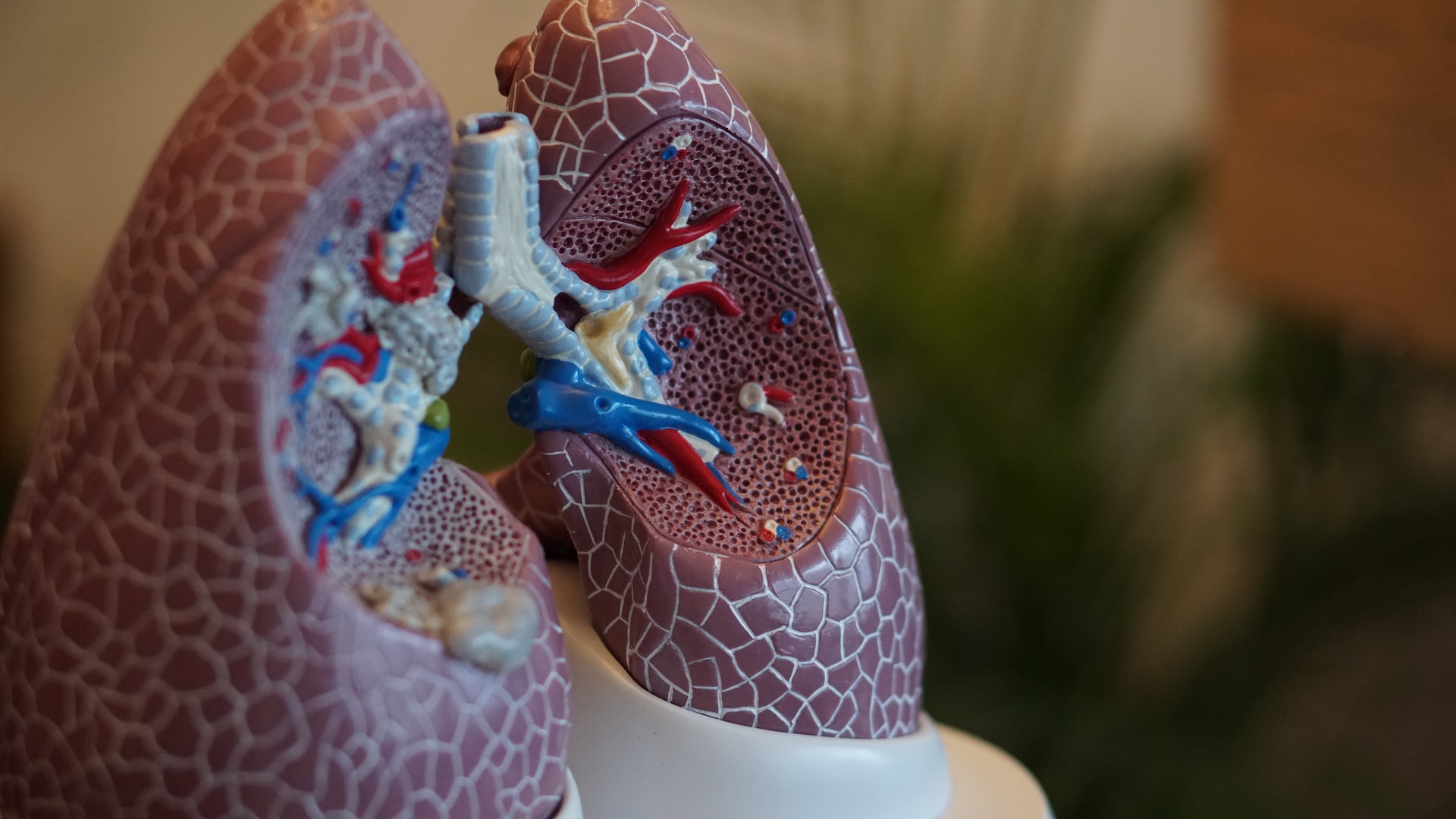
Pleural cavity is the space between the visceral pleura, which is adjacent to the lungs, and the parietal pleura. It contains serous fluid in small amounts. The fluid keeps the surface of the lungs moist all the time, making it easier for the lungs to move when breathing.
Taking into account the mechanism of formation and location of pneumothorax, the following types of pneumothorax are distinguished:
Closed pneumothorax: Closed pneumothorax occurs when a small amount of air enters the pleural cavity without a traumatic or iatrogenic cause![]() . The air is spontaneously absorbed over time. If necessary, a puncture can be performed to remove the accumulated air.
. The air is spontaneously absorbed over time. If necessary, a puncture can be performed to remove the accumulated air.
Open pneumothorax: Open pneumothorax occurs when air enters the pleural cavity through an opening in the chest or bronchi caused by penetrating chest wound![]() . The same space allows air to escape from the lungs. This condition poses a serious risk to the patient's life as it impairs respiratory capacity by compromising the function of one lung.
. The same space allows air to escape from the lungs. This condition poses a serious risk to the patient's life as it impairs respiratory capacity by compromising the function of one lung.
Tension pneumothorax: Tension pneumothorax occurs due to trauma or gunshot wounds. In this condition, each breath drawn in by the patient introduces air into the pleural cavity. However, this air cannot escape through the same pathway it entered. With each subsequent inhalation, more air accumulates in the confined space, causing pressure within the pulmonary cavity and impeding organ expansion. Consequently, pneumothorax continues to expand and ultimately causes lung collapse![]() .
.
Understanding pneumothorax and its various factors is crucial for accurate diagnosis and treatment. By categorizing it based on specific factors, healthcare professionals can gain a deeper understanding of the condition's mechanisms and provide appropriate care. Additionally, dividing pneumothorax into small and large sizes helps determine its severity and tailor treatment methods accordingly. This enables doctors to intervene effectively and ensure optimal patient outcomes.
Small pneumothorax: In a small pneumothorax, the space between the chest wall and the pleura is less than 1 inch. This is considered a minor level of pneumothorax, where there is only a limited amount of excess air in the pleural cavity.
Large pneumothorax: The distance between the chest wall and pleura may be more than 1 inch. It is a more advanced level of pneumothorax, where the excess air in the pleural cavity is significant.
Pneumothorax can occur for various reasons. Most often, pneumothorax occurs spontaneously through rupture of the emphysematous cyst or alveoli close to the pleura. Such a situation can occur in healthy people without lung disease and in the course of lung and bronchial diseases. Depending on the factor that causes pneumothorax, the following types are distinguished:
It is an pneumothorax that occurs as a complication after various medical procedures![]() done in the chest area. Examples include cardiac surgery or central venipuncture. Cardiac surgery is a medical field that includes the surgical treatment of diseases of the heart and blood vessels. For example, cardiac surgery for coronary artery disease and valvular heart defects is the standard procedure.
done in the chest area. Examples include cardiac surgery or central venipuncture. Cardiac surgery is a medical field that includes the surgical treatment of diseases of the heart and blood vessels. For example, cardiac surgery for coronary artery disease and valvular heart defects is the standard procedure.
On the other hand, a central puncture is a procedure where a catheter is inserted into a large main vein through a blood vessel. This type of puncture is typically done in the subclavian vein. It's important to note that when an abscess is drained into the pleural cavity, it can cause pneumothorax. This occurs when air from the abscess cavity enters the pleural space through the lung.
The exact incidence of primary spontaneous pneumothorax is uncertain![]() . While the causes can vary, cigarette smoking is one of the primary risk factors. Smoking causes emphysematous foci in the lungs, which increases the risk of pneumothorax. Rupture of subpleural emphysematous alveoli in the lungs can occur during severe coughing.
. While the causes can vary, cigarette smoking is one of the primary risk factors. Smoking causes emphysematous foci in the lungs, which increases the risk of pneumothorax. Rupture of subpleural emphysematous alveoli in the lungs can occur during severe coughing.
People with pneumothorax often have weakened alveoli, which can rupture due to violent coughing. Air then enters the pleural space, causing pneumothorax. A form of pneumothorax also occurs during menstruation which can occur in women of childbearing age.
Symptoms are similar to ordinary pneumothorax but tend to follow a cyclic pattern combined with menstruation. It is associated with a small amount of tissue from the endometrium that enters and nests in the pleura. There is also secondary spontaneous pneumothorax that can occur in various lung diseases.
It is an pneumothorax that occurs due to trauma![]() to the chest, especially where there is damage to the outer shell. An example is a broken rib, which can puncture the skin and cause pneumothorax. Pneumothorax can also occur as a result of trauma to the chest.
to the chest, especially where there is damage to the outer shell. An example is a broken rib, which can puncture the skin and cause pneumothorax. Pneumothorax can also occur as a result of trauma to the chest.
The most common injuries causing pneumothorax occur in traffic accidents, falls from heights, crushing, or being stabbed with a sharp object. It is events that often require surgical intervention. In the case of pneumothorax, drainage is recommended to drain excess air from the pleura. Contour deformity usually requires surgical treatment to stabilize the fragments with specialized instrumentation.
The severity of symptoms in pneumothorax depends on the type of condition. For instance, in cases of spontaneous pneumothorax, pneumothorax can be symptomless. Typically, the absence of symptoms indicates a small pneumothorax. As the size of the pneumothorax increases, symptoms become more severe. If a significant amount of air enters the pleural cavity, patients may experience intense pain.
The signs of pneumothorax are distinctive and recognizable even to individuals without medical expertise, prompting them to seek emergency care when necessary. General symptoms of pneumothorax include:
Chest pain: Individuals commonly experience sudden and intense chest pain. This pain may become more severe after coughing, engaging in strenuous physical activity, or experiencing chest trauma. It is often described as a sensation of pressure or tightness in the chest area![]() . This type of pain is one of the most distinctive symptoms of pneumothorax. Additionally, the pain may radiate to other parts of the body, including the neck, shoulder, and abdomen.
. This type of pain is one of the most distinctive symptoms of pneumothorax. Additionally, the pain may radiate to other parts of the body, including the neck, shoulder, and abdomen.
Shortness of breath: One common symptom of pneumothorax is shortness of breath, which typically develops rapidly and worsens as the condition progresses. Patients may experience difficulty breathing![]() and compensate by taking rapid breaths to try to compensate for the impaired gas exchange. This can cause significant feelings of breathlessness. Additionally, individuals with pneumothorax may notice an increase in their chest circumference.
and compensate by taking rapid breaths to try to compensate for the impaired gas exchange. This can cause significant feelings of breathlessness. Additionally, individuals with pneumothorax may notice an increase in their chest circumference.
Tachycardia: Tachycardia, or an accelerated heart rate![]() , often accompanies a pneumothorax. When there is excess air in the pleural cavity, it irritates the pleural membrane and can lead to an increased heart rate. If air freely flows into and out of the pleural cavity through a chest wall opening, it can cause dangerous mediastinal pendulums and potentially result in reflex cardiac arrest.
, often accompanies a pneumothorax. When there is excess air in the pleural cavity, it irritates the pleural membrane and can lead to an increased heart rate. If air freely flows into and out of the pleural cavity through a chest wall opening, it can cause dangerous mediastinal pendulums and potentially result in reflex cardiac arrest.
Sweating: Excessive sweating is a common symptom experienced by patients with pneumothorax. This excessive sweating is a result of the physical stress that accompanies the condition, as well as the body's response to stress and hypoxia. The natural process of sweat secretion helps regulate and maintain average body temperature.
Pallor of the skin: Due to impaired circulation and gas exchange, patients with pneumothorax may exhibit pallor of the skin coverings. Pallor is the result of tissue hypoxia and restricted blood flow. In severe cases, hypoxia can lead to cyanosis of the face and neck.
Weakness: As a pneumothorax worsens, patients may experience growing weakness throughout their body. This is due to the lack of oxygen, which affects overall bodily function. In extreme cases of hypoxia, fainting may also occur.

The level of danger associated with pneumothorax depends on the type and size of the condition. Tension pneumothorax, for example, is a highly dangerous and immediately life-threatening situation![]() . For other types of pneumothorax, such as minor spontaneous cases, the critical factor is the size or collapse level of the lung. These cases often require observation or a simple puncture or pleural drainage procedure. Regardless of its type, pneumothorax is a potentially life-threatening condition that requires prompt medical intervention.
. For other types of pneumothorax, such as minor spontaneous cases, the critical factor is the size or collapse level of the lung. These cases often require observation or a simple puncture or pleural drainage procedure. Regardless of its type, pneumothorax is a potentially life-threatening condition that requires prompt medical intervention.
Pneumothorax can pose a life-threatening risk to patients and requires prompt treatment. If left untreated, it can lead to acute respiratory failure and potentially result in death. However, small pneumothoraxes are easily treatable, and the condition is generally curable![]() . In more severe cases where there is significant pneumothorax, doctors may opt for a procedure called pleural aspiration using a Venflon needle inserted into the pleura through the chest wall. This method presents greater seriousness and practicality under certain circumstances.
. In more severe cases where there is significant pneumothorax, doctors may opt for a procedure called pleural aspiration using a Venflon needle inserted into the pleura through the chest wall. This method presents greater seriousness and practicality under certain circumstances.
A medical interview with the patient may be sufficient to diagnose pneumothorax with characteristic symptoms and large size. If the symptoms are less typical, it is necessary to conduct numerous tests to confirm or exclude the presence of pneumothorax. Depending on the clinical situation, the doctor may perform one or more tests to confirm or exclude pneumothorax. Among the tests necessary in the diagnosis of pneumothorax are:
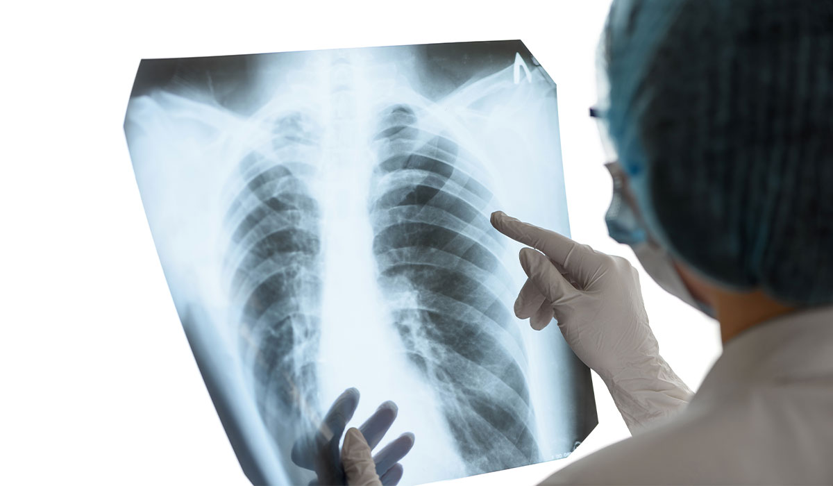
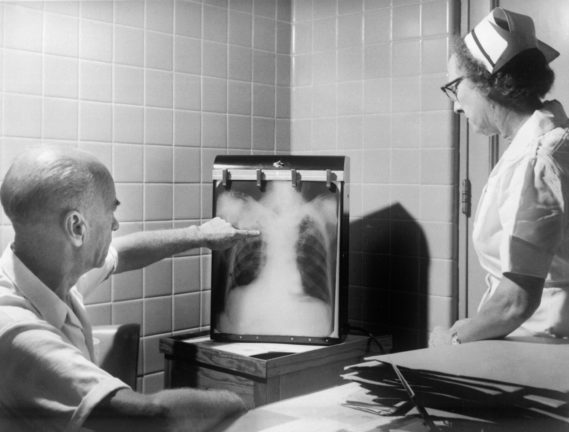
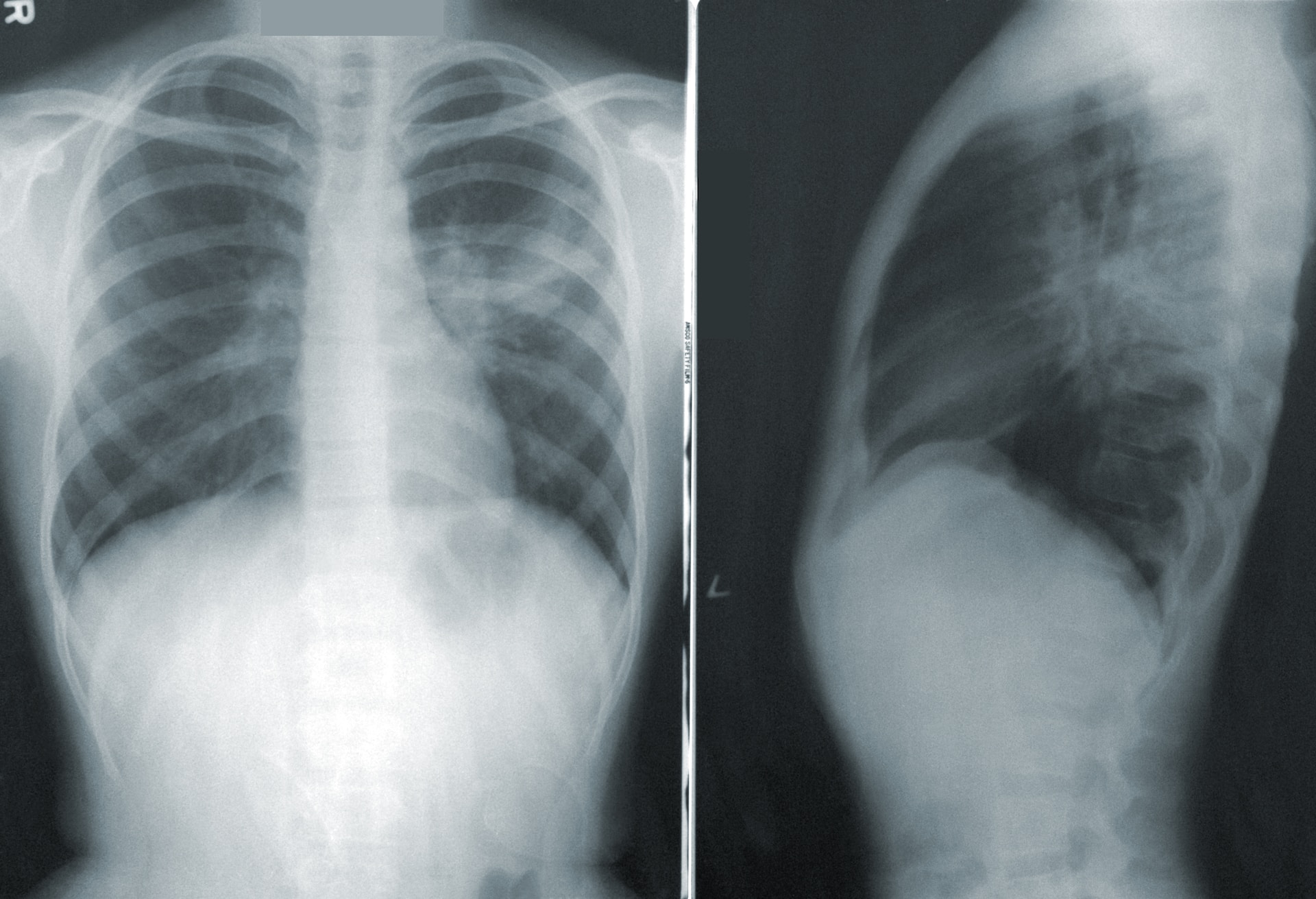
Chest X-ray: A chest X-ray is the main diagnostic test for pneumothorax. It allows doctors to assess the condition of the lungs and determine the location and size of air in the pleural cavity. X-rays are also performed after accidents or chest trauma to monitor the progression of pneumothorax over time, often with a series of images taken at regular intervals.
Chest ultrasound: Chest ultrasound is a valuable tool for diagnosing various chest lesions and injuries, especially in emergency situations. It allows doctors to quickly assess the condition of the pleural cavity and identify any significant gas concentrations.
CT scan: CT scan of the chest is a good examination for a thorough morphological evaluation of the lungs and other structures in the chest. It is a more sensitive test than an X-ray, making it possible to see changes in trauma patients whose pneumothorax is initially invisible.
Cardiac electrocardiography: An increased heart rate is a common symptom accompanying pneumothorax so an ECG may be helpful in some cases. It is an auxiliary test that can be used for a sizeable unilateral pneumothorax. The recording then shows conduction abnormalities.
Blood tests: Additionally, healthcare providers use blood gasometry![]() to measure the levels of respiratory gases and maintain the balance of acid and base in the blood. Pulse oximetry is another non-invasive method that assesses oxygen levels in the blood, providing valuable information about respiratory capacity.
to measure the levels of respiratory gases and maintain the balance of acid and base in the blood. Pulse oximetry is another non-invasive method that assesses oxygen levels in the blood, providing valuable information about respiratory capacity.
Determining the best treatment method depends on various factors, including the patient's specific condition and the size of the collapsed lung. The severity of symptoms is also taken into consideration. The primary goal of medical treatment is to remove air from the pleural cavity, reflate the collapsed lung, and prevent future recurrence.
Small pneumothorax: For a small pneumothorax, the patient can receive treatment while staying in bed. This includes taking pain medication and doing breathing exercises. In non-life-threatening cases where the patient is experiencing a small pneumothorax for the first time, it is recommended to rest, do breathing exercises, and receive oxygen therapy. Typically, without medical intervention, the air trapped in the chest will be absorbed within a few days.
Large pneumothorax: In cases of a large pneumothorax, prompt medical attention is necessary. Oxygen should be administered if there is an immediate threat to life. If it's a unilateral pneumothorax and the lung and major blood vessels are under pressure, it may be necessary to puncture the pleura and rapidly drain the gases. On the other hand, bilateral pneumothorax involves drainage of the pleural cavity, which should be started on the side with the larger pneumothorax.

Considering the size of the pneumothorax and the severity of the patient's condition, the following treatment techniques are used:
Pain therapy: In pneumothorax cases, pain medications are often used to relieve symptoms. In cases of a small pneumothorax, recovery can be achieved through the use of pain medication, rest, and prescribed breathing exercises. Severe cases may require additional painkillers to help alleviate discomfort for the patient.
Oxygen therapy: Oxygen therapy is a treatment used to address respiratory failure. It involves increasing the amount of oxygen in the air that a patient inhales. Oxygen therapy![]() is necessary in most cases of pneumothorax. However, there are contraindications to this treatment method, including pregnant women should not use oxygen therapy.
is necessary in most cases of pneumothorax. However, there are contraindications to this treatment method, including pregnant women should not use oxygen therapy.
Asherman's Chest Seal: The Asherman's Chest Seal![]() is a dressing specifically designed for open chest wounds. It features a built-in one-way valve that allows excess air and blood to escape while preventing them from re-entering the chest cavity. This dressing is particularly effective in cases where there is an open pneumothorax, ensuring proper airflow regulation and promoting healing.
is a dressing specifically designed for open chest wounds. It features a built-in one-way valve that allows excess air and blood to escape while preventing them from re-entering the chest cavity. This dressing is particularly effective in cases where there is an open pneumothorax, ensuring proper airflow regulation and promoting healing.
Air aspiration puncture: Air aspiration puncture![]() is a procedure used to remove trapped air in the pleural cavity when there is a subpleural rupture. It involves inserting a needle into the hole and aspirating the air. This minimally invasive technique aims to alleviate lingering air pockets in the pleural space. It is applicable only in first-time cases. If a pneumothorax recurrence occurs, different methods are used to treat the condition.
is a procedure used to remove trapped air in the pleural cavity when there is a subpleural rupture. It involves inserting a needle into the hole and aspirating the air. This minimally invasive technique aims to alleviate lingering air pockets in the pleural space. It is applicable only in first-time cases. If a pneumothorax recurrence occurs, different methods are used to treat the condition.
Pleural cavity drainage: Pleural cavity drainage![]() is a procedure done under local anesthesia. It involves inserting a thin drain tube into the pleura through the intercostal space. In cases of large pneumothorax, a drain is secured to the skin with a suture and connected to a system that allows air to escape from the pleura. This drain remains in place for several days.
is a procedure done under local anesthesia. It involves inserting a thin drain tube into the pleura through the intercostal space. In cases of large pneumothorax, a drain is secured to the skin with a suture and connected to a system that allows air to escape from the pleura. This drain remains in place for several days.
Operative treatment: In more severe cases, when other treatment methods have not been effective, surgical intervention may be necessary to achieve the desired results. Indications for surgery include recurrent pneumothorax. Pleurodesis, which involves treatment leading to overgrowth of the pleural cavity, and pleurectomy, which requires removal of the mural pleura, are then used. The newest surgical treatment option is Video Associated Thoracoscopic Surgery![]() . The method removes lesions in the lung parenchyma and pleura using special miniature instruments inserted into the chest through two small incisions.
. The method removes lesions in the lung parenchyma and pleura using special miniature instruments inserted into the chest through two small incisions.
After undergoing pneumothorax treatment, a safe lifestyle is recommended to prevent pneumothorax recurrence. Post-therapeutic principles are as follows:

No flying: The pressure difference during flight can cause the air pockets in the pleural space to expand, consequently worsening the existing pneumothorax and the patient's condition. For this reason, it is discouraged to fly aboard cruise passenger aircraft for this condition. However, airline services can be used for at least two weeks![]() after favorable treatment.
after favorable treatment.
No diving: Note that many respiratory diseases are absolute contraindications to diving. Including a history of pneumothorax is a complete and lifelong contraindication to diving![]() . Once you have a pneumothorax, you should never dive to great depths, bungee jump, or skydive. Thus, the high-pressure differential can pose a significant danger to people after pneumothorax.
. Once you have a pneumothorax, you should never dive to great depths, bungee jump, or skydive. Thus, the high-pressure differential can pose a significant danger to people after pneumothorax.
Smoking cessation: It is strongly recommended that all patients quit smoking, as nicotine use increases the likelihood of pneumothorax recurring. Pneumothorax is more prevalent in younger individuals and smoking![]() significantly contributes to this risk. Smoking leads to the development of air-filled pockets, known as pneumothorax bubbles, within the lungs.
significantly contributes to this risk. Smoking leads to the development of air-filled pockets, known as pneumothorax bubbles, within the lungs.
Restriction of physical activity: Once pneumothorax has been treated, this includes limitation of physical activity. Exemption from strenuous exercise should last a minimum of two weeks after treatment. During this time, special rehabilitation exercises to improve lung function will be the best choice.
After the initial episode of pneumothorax; some individuals may experience a recurrence![]() where air accumulates in the pleural cavity once again. Recurrences can occur with closed and open pneumothorax cases, which are standard. There are various causes for recurrent pneumothorax, including:
where air accumulates in the pleural cavity once again. Recurrences can occur with closed and open pneumothorax cases, which are standard. There are various causes for recurrent pneumothorax, including:
Regular monitoring and preventive strategies should be implemented to prevent recurring pneumothorax. This includes avoiding risk factors and scheduling regular medical check-ups to detect and treat any potential recurrences early on. Individuals with a history of pneumothorax should receive ongoing medical care due to the risk of recurrence.
Pneumothorax primarily affects males, making it rare in pregnant women. However, there are occasional cases where pregnant women can develop pneumothorax. In these instances, the changes in a pregnant woman's respiratory system can pose complications and risks to both the mother and fetus—pneumothorax during pregnancy often results in hypoxia![]() , characterized by chest pain and shortness of breath.
, characterized by chest pain and shortness of breath.

Proper diagnosis is crucial because the symptoms can sometimes be mistaken for pregnancy-related difficulty in breathing. Although the use of radiologic studies is limited in such cases, a thorough medical history can aid in determining the correct diagnosis.
Ultrasound, which poses no risks to the mother or fetus, is commonly used during pregnancy for procedures like amniocentesis and fetal blood transfusion. It can also help detect pregnancy itself. Similarly, MRI has been safely utilized to address lung issues in pregnant individuals without harming both mother and baby.
When it comes to treating pneumothorax during pregnancy, several approaches are available. As we know, pregnant women have a higher risk of experiencing pneumothorax, and treating it using conservative measures is essential. In most cases, conservative methods and tube thoracostomy yield satisfactory results.
However, for instances where prolonged air leakage or recent recurrences occur, video-assisted thoracoscopic surgery (VATS) may be necessary. Regarding delivery options for pregnant women with pneumothorax, a cesarean section under epidural anesthesia is considered a safe choice.
Pneumothorax is a condition where air builds up in the pleural space, which is the area between the lung's internal pleura and the chest wall's external pleura. Air aspiration puncture is a procedure used to release trapped air when there is a subpleural rupture. It involves inserting a needle into the hole and removing the air. This minimally invasive technique aims to relieve lingering pockets of air in the pleural space.
When there is excess air in the pleural cavity, it causes the two layers to separate, putting pressure on the lung and hindering its normal respiratory function. This commonly leads to chest pain and shortness of breath. Pneumothorax is a serious condition that can put your life and health at risk. There are different types of pneumothorax categorized by their size or how they are formed.
The symptoms experienced and their severity depend on the specific type of pneumothorax. However, some cases may not show any symptoms at all. In severe cases, distinct symptoms would help diagnose the condition correctly, while additional tests may be necessary for smaller pneumothorax cases. The primary goal when treating pneumothorax is to remove air from the pleural cavity, re-expand the collapsed lung, and restore normal respiratory function.
Treatment options vary based on the type and severity of pneumothorax symptoms. It's important to note that after receiving treatment for pneumothorax, caution should be exercised to avoid actions that could worsen the condition or contribute to a recurrence. Although treatable, recurrences can still happen with this condition; therefore, vigilance is required even after treatment has been administered successfully.
Pneumothorax occurs more frequently in men but can also affect women (including those who are pregnant). If pregnant women experience pneumothorax. treatment should be administered immediately, as pneumothorax can lead to pregnancy complications.