X-Ray is a form of electromagnetic radiation widely used in medicine and allows for beneficial diagnostic imaging studies. This diagnostic test takes advantage of the X-ray's ability to penetrate many objects, including the human body.
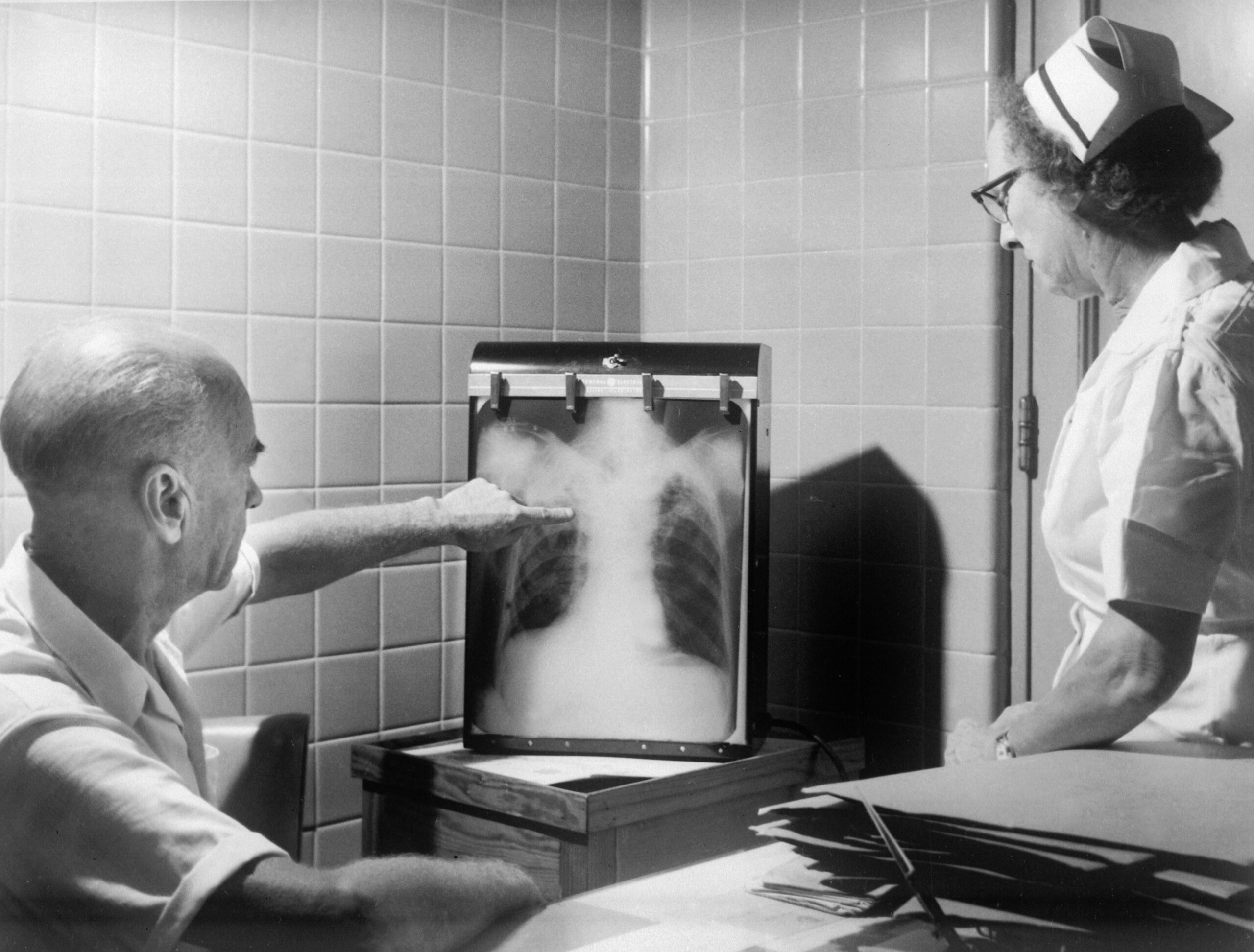
X-rays pass through the body and are absorbed in varying amounts. The procedure makes generating images of tissues and areas inside the body possible. There are different types of X-ray detectors. X-ray images are called radiographs. Patients are placed between the X-ray source and detector during the examination.
X-ray examination requires the observance of certain principles. X-rays are widely used in medicine in many different situations, facilitating the diagnosis of many diseases. X-rays have also been found to be helpful in medicine for cancer treatment. X-rays can react with atoms and cells, which can cause specific hazards. The human body's cells can trigger repair processes to eliminate the adverse effects of X-rays. However, overexposure to X-rays can carry adverse effects. Specific contraindications to X-ray examination must be observed.

X-Ray was discovered in Germany by Wilhelm Conrad Röntgen![]() , who performed experiments in 1895
, who performed experiments in 1895![]() . He used Crookes tubes, devices usually used by scientists to visualize electron streams. Wilhelm noticed that the radiation was passing through black cardboard. Noticing the penetration through objects was crucial and led to systematic research into a new type of radiation. Wilhelm Conrad Röntgen named the radiation ‘X.' The first X-ray image showed a cross-section of Wilhelm's wife's hand.
. He used Crookes tubes, devices usually used by scientists to visualize electron streams. Wilhelm noticed that the radiation was passing through black cardboard. Noticing the penetration through objects was crucial and led to systematic research into a new type of radiation. Wilhelm Conrad Röntgen named the radiation ‘X.' The first X-ray image showed a cross-section of Wilhelm's wife's hand.
After this remarkable discovery, the scientist presented the first paper on X-rays. The man was awarded the Nobel Prize in Physics![]() for his discovery. Today, X-rays are widely used by doctors and specialists. Its popularity has grown due to its commercial approach, which has resulted in the X-ray being used in medical facilities worldwide. Several companies were responsible for the commercial implementation of radiation. The production of X-ray tubes has been commercially successful, and some companies are still in operation today.
for his discovery. Today, X-rays are widely used by doctors and specialists. Its popularity has grown due to its commercial approach, which has resulted in the X-ray being used in medical facilities worldwide. Several companies were responsible for the commercial implementation of radiation. The production of X-ray tubes has been commercially successful, and some companies are still in operation today.

X-rays have a wide range of applications. It is a quick and readily available examination that, using a low dose of radiation, enables an image of a particular body area. The duration of the examination depends on the number of projections required. X-ray examination is essential in diagnosing injuries, especially fractures, but it also helps assess the condition of organs. In addition to the diagnostic process, radiation can be used for therapeutic purposes. Examples of X-ray applications include:
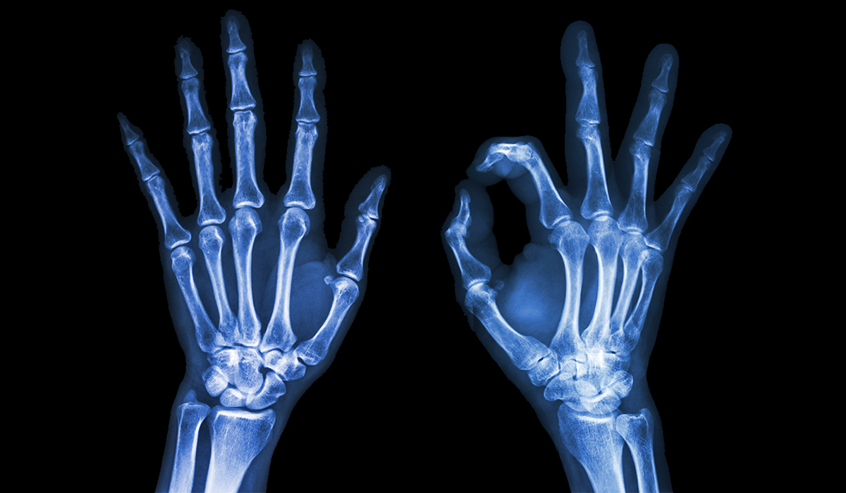
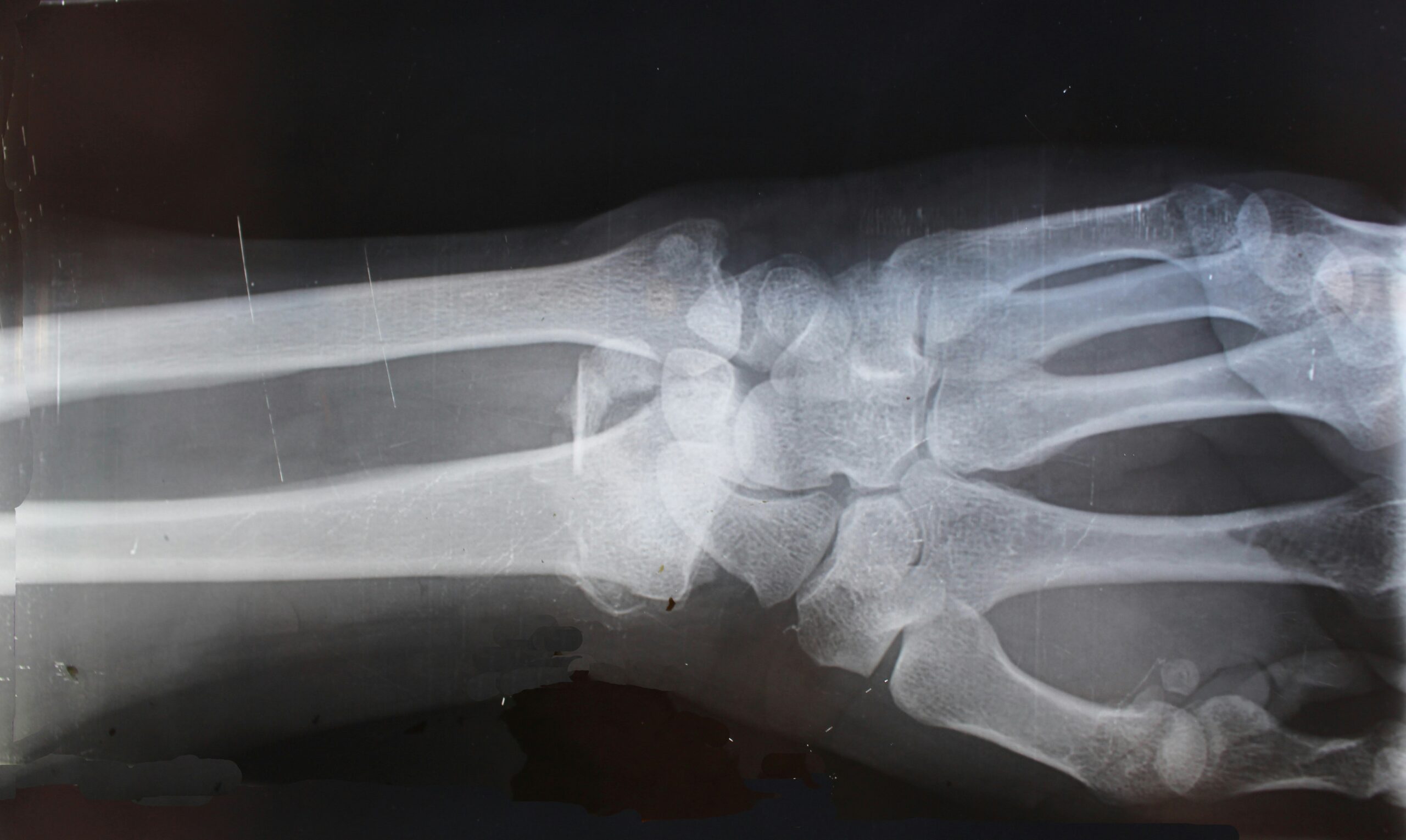
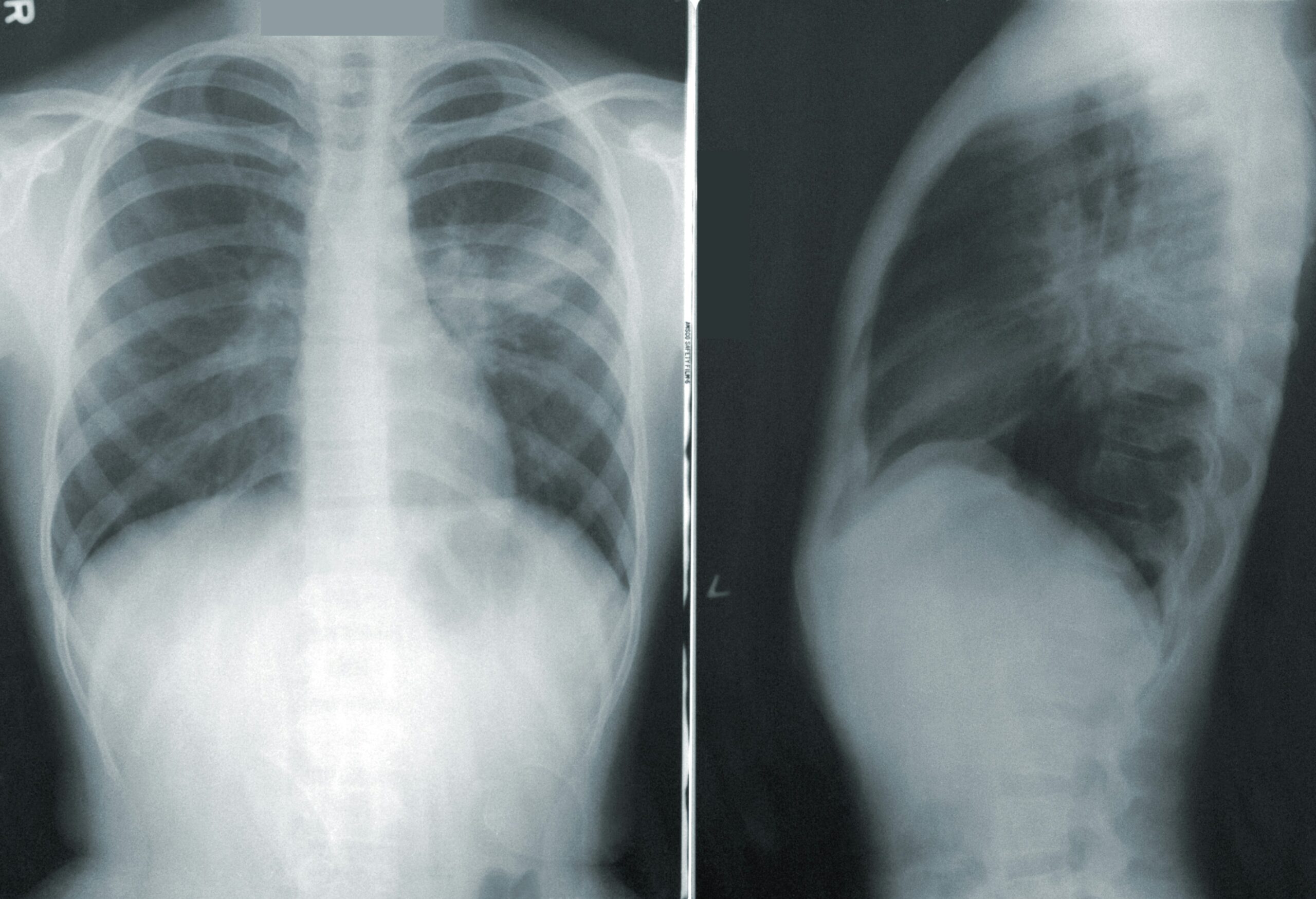
The procedure is the basis of diagnostics that detects many ailments, such as bone fractures, tumors, and foreign bodies inside the human body. Depending on the case, X-rays can be taken on different body areas. X-ray photography of bones and joints can detect injuries and degeneration. Another example is an X-ray of the gastrointestinal tract, which is helpful in cases of perforations in this area.
X-rays and CT scans are ionizing radiation-based examinations that differ from each other in several aspects. CT is a combination of X-ray technology and computer processing. The assessment generates a series of cross-sectional images of the body that can be combined into a three-dimensional X-ray image. CT is characterized by the higher resolution of the images obtained and the greater ability to differentiate tissues and image pathological conditions. X-ray versus CT also differs in the examination procedure.
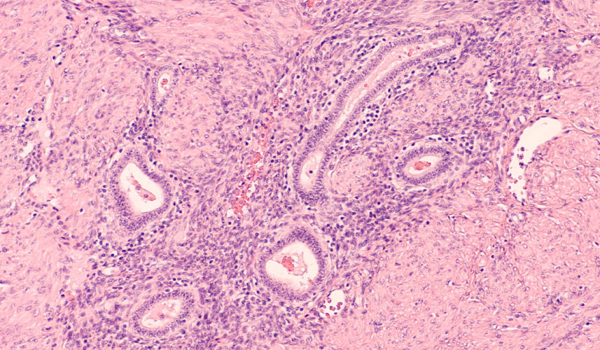
Mammography is the name for an examination using X-rays to detect and diagnose cancers in the breast area. Tumors and small calcium fragments can be seen on X-rays, which is important because microcalcifications can sometimes indicate the presence of a particular type of cancer. Mammography has a high sensitivity and may work better than ultrasound. Mammography performed regularly in women is a recommended screening test.
X-rays can also be combined with a fluorescence screen. This type of examination is performed to obtain real-time body movement images. The procedure facilitates the diagnostic process by following the path of the contrast agent that has been administered to the patient. The fluorescence screen can show the movement of the beating heart and the blood flow to the heart muscle. The procedure helps assess blood vessels and organ function. Fluoroscopy is practical not only in diagnosis but also in therapy, including image-guided surgery. The examination helps remove foreign bodies from deep tissues.
Specialists use high-energy radiation to destroy cancer cells, among other things. The high frequency of the waves causes ionization in the tissue. The radiation destroys tumors by damaging the DNA of the cancer cells. Higher doses of radiation are used for therapeutic purposes than for diagnosis. Radiation therapy for oncology is one of the three main treatments for patients with malignant tumors. It is used alone or integral to combination treatment with surgery, chemotherapy, and other systemic cancer treatments.
X-rays are under classical conditions generated by an X-ray tube. Such a tube is constructed from a vacuum bulb and the physical processes taking place within it allow X-ray beams to be produced. The examination involves X-raying a patient with a beam of X-rays to obtain an image of a particular body area.
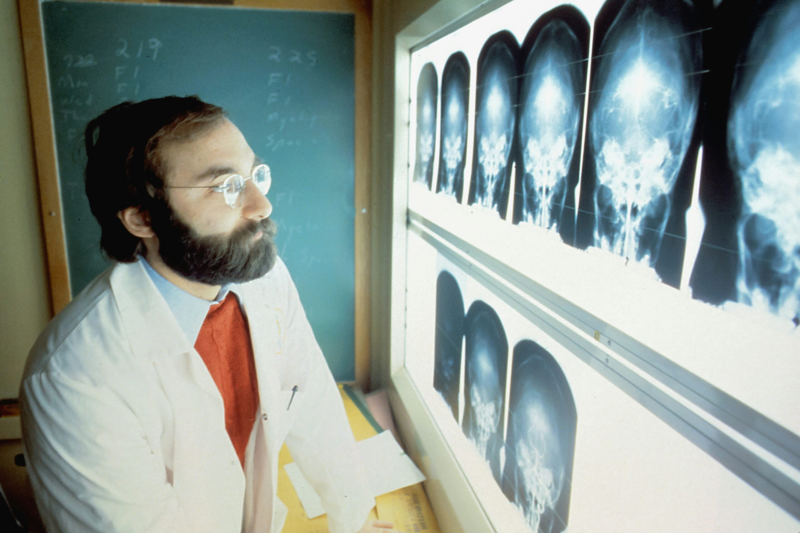
The duration of the examination depends on the number of pictures taken and how the patient's body is positioned. The patient is positioned between an apparatus that records the radiological image and a device that emits X-rays. The device can be arranged differently, depending on the area of the body being examined. The X-ray tube can be suspended above the bed on which the patient is lying. The examination can also be performed in a standing or sitting position. Most medical laboratories are equipped with digital X-ray machines, and the X-ray results are given to the patient after the examination.
It would be best to prepare for the X-ray examination according to your doctor's instructions. This particularly applies if contrast material is to be administered. On the day of the X-ray examination, you should usually be fasting. Also, for imaging examinations, you should remove any metal items within the study area, i.e., jewelry and other accessories. Your preparation will maximize the effectiveness and accuracy of the examination.
The X-ray procedure is also extended for examinations that require administering shadowing material to the patient. Contrast materials are used in radiology. These are chemical substances introduced by various routes into the body. Contrast materials allow an increase in the contrast of the images obtained during radiological examinations by increasing the brightness differences. This method translates into better visualization of the body structures under examination and, in particular, better visualization of pathological changes needed for diagnosis.
Modern contrast agents do not usually cause side effects, but this is sometimes possible. When a preparation is introduced into the body, it can cause transient effects. If side effects do occur, they are usually of minor intensity. However, severe or life-threatening reactions have also been described. It is, therefore, important to exercise caution and follow current guidelines related to the use of contrast agents.
Different contrast agents are used for different examination techniques. The materials can also be administered in different ways, which include:
In some cases, oral administration of an agent in the form of a tablet or liquid that enters the patient's body by swallowing is sufficient. Substances are used in this way for gastrointestinal studies, for example. Among others, substances such as barium sulfate![]() suspensions are used orally. Barium sulfate is insoluble in water and is not absorbed from the gastrointestinal tract. A contrast agent administered orally may cause side effects such as diarrhea. Symptoms disappear when the contrast agent is emptied from the intestine.
suspensions are used orally. Barium sulfate is insoluble in water and is not absorbed from the gastrointestinal tract. A contrast agent administered orally may cause side effects such as diarrhea. Symptoms disappear when the contrast agent is emptied from the intestine.
Many contrast materials are administered intravenously by injecting the substance into a vein or artery. Such purposes include using iodine![]() contrast agents excreted through the kidneys. Materials can be administered directly into the gastrointestinal tract, urinary tract, biliary tract, reproductive tract, and spinal canal. Intravenous administration of iodine is most commonly used in CT scans. Symptoms like pain and redness at the site of contrast administration may occur with intravenous administration.
contrast agents excreted through the kidneys. Materials can be administered directly into the gastrointestinal tract, urinary tract, biliary tract, reproductive tract, and spinal canal. Intravenous administration of iodine is most commonly used in CT scans. Symptoms like pain and redness at the site of contrast administration may occur with intravenous administration.
An enema is also used in X-ray examinations, which allows rectal administration of contrast substances. The rectal method also uses a barium preparation, which helps the walls of the colon to be evenly covered. Contrast-enhanced colon examinations are performed in suspected proliferative processes, intestinal obstruction, and inflammatory bowel disease. A side effect of rectal administration may be diarrhea or stool coloring.
X-ray examinations are generally relatively safe and can diagnose life-threatening conditions, so their usefulness often outweighs the risks. However, overexposure to X-rays can carry adverse effects. This type of examination produces ionizing radiation that can damage living tissue.
The more significant health risks include the greater strength of the energy used and the exposure. The effects of radiation also vary depending on the location of the tissue or organ. Shortened life expectancy, radiation-induced malignancies![]() , and genetic damage
, and genetic damage![]() are common biological radiation risks. However, X-ray risks are generally low. Some groups are at higher risk of harmful X-ray radiation, and these include:
are common biological radiation risks. However, X-ray risks are generally low. Some groups are at higher risk of harmful X-ray radiation, and these include:

Choosing the safest imaging method for pregnant patients is essential to medicine. The uniqueness of pregnancy is that exposure of the fetus to radiation is a significant risk factor that can pose a threat to health and development. High exposure to X-rays can be harmful to pregnant women because the fetus retains some of the radiation. Examples of radiation effects on the fetus include various disease entities and even fetal death![]() . Fetuses are more exposed to radiation than adult patients because they have a rapidly developing cellular system.
. Fetuses are more exposed to radiation than adult patients because they have a rapidly developing cellular system.
Higher levels of radiation increase the likelihood of malformations![]() . The health effects of radiation vary according to gestational age. The first trimester of pregnancy is the most sensitive period for X-ray examination. Therefore, it is often necessary to perform a pregnancy test before a woman undergoes the examination. Imaging diagnosis of pregnant women must, therefore, be carried out with great care and safety. In pregnant women, doctors, therefore, prefer to use tests that do not use radiation, such as MRI or ultrasound.
. The health effects of radiation vary according to gestational age. The first trimester of pregnancy is the most sensitive period for X-ray examination. Therefore, it is often necessary to perform a pregnancy test before a woman undergoes the examination. Imaging diagnosis of pregnant women must, therefore, be carried out with great care and safety. In pregnant women, doctors, therefore, prefer to use tests that do not use radiation, such as MRI or ultrasound.
Another group that is more exposed to the harmful effects of X-ray tubes is children. Children are more likely to develop radiation-induced cancer. X-ray examinations early in life can result in several times the risk of harmful effects compared to adults. Children also have a higher exposure to the effects of ionizing radiation in the form of genetic mutations. The long-term side effects of radiation are, therefore, a greater risk for children. It is, therefore, essential to use dose limits that are lower for children. There are also alternative methods of diagnosis, such as ultrasound and MRI, which are becoming increasingly available.
X-rays, are among the electromagnetic radiations used in medicine. Radiation was discovered by Wilhelm Conrad Roentgen in 1895, which initiated the sale of X-ray tubes used by doctors. X-rays pass through matter that is not transmitted by light. The X-ray examination makes it possible to visualize the inside of the human body in an image. There are different X-ray examinations; other body areas can be examined.
X-rays are also used for therapeutic purposes to treat cancer. The procedure for this type of examination is not complicated, and X-ray examinations are quick and easily accessible. To increase accuracy, specialists also use contrast materials. However, an X-ray examination is associated with risks related to the harmful effects of ionizing radiation.