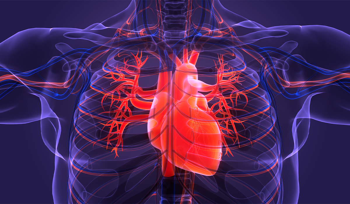
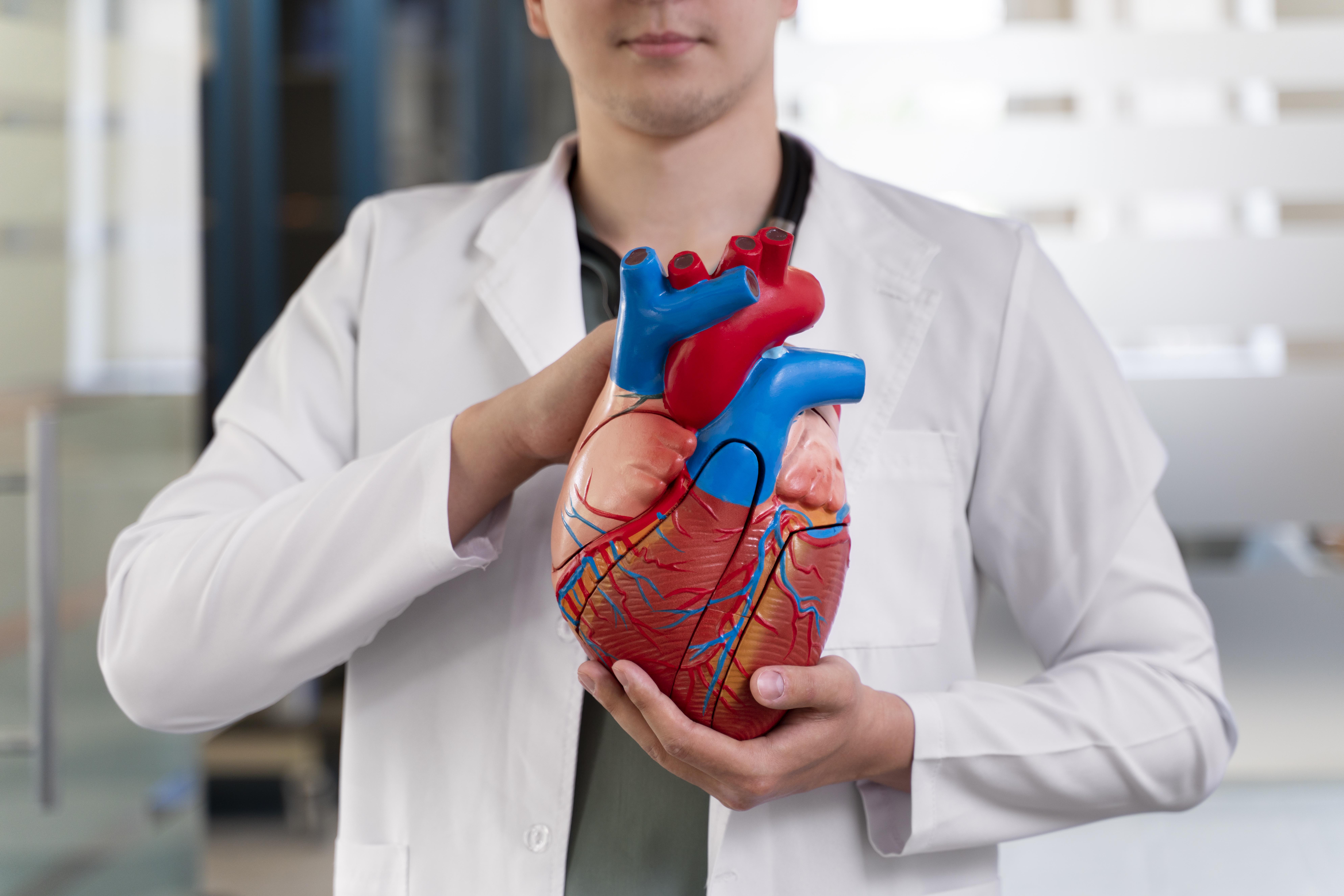
Angina is not a separate disease but rather a manifestation of ischemic heart disease. The heart muscle is supplied with blood by the coronary arteries. Blood supplies the heart with the necessary energy substances and oxygen.
In healthy, young people, the heart has a certain coronary reserve, which means that in the event of increased exercise, the heart may receive up to several times more blood than at rest. Coronary heart disease most often develops due to atherosclerosis. It causes the narrowing of the coronary vessels, thus impeding blood flow. Lack of coronary reserve causes angina pain in cases of stress or increased exertion.
We distinguish between stable![]() and unstable angina
and unstable angina![]() . Stable angina should subside within 5 minutes of stopping exercise or taking a vasodilator. Stable angina usually requires drug treatment and lifestyle modifications. Coronary angiography – as an invasive treatment – is often not necessary. However, unstable angina is one of the acute coronary syndromes.
. Stable angina should subside within 5 minutes of stopping exercise or taking a vasodilator. Stable angina usually requires drug treatment and lifestyle modifications. Coronary angiography – as an invasive treatment – is often not necessary. However, unstable angina is one of the acute coronary syndromes.
The difference between angina and a heart attack is that in the case of angina, necrosis of the heart muscle cells has not yet occurred. However, during an infarction, ischemic cells die.
Angina![]() is most often caused by atherosclerosis of the coronary arteries. These vessels bring blood to the heart and are therefore accountable for supplying it with oxygen and nutrients. The narrowing of the diameter of coronary arteries occurs due to the formation of atherosclerotic plaque. The following contribute to its development, among others:
is most often caused by atherosclerosis of the coronary arteries. These vessels bring blood to the heart and are therefore accountable for supplying it with oxygen and nutrients. The narrowing of the diameter of coronary arteries occurs due to the formation of atherosclerotic plaque. The following contribute to its development, among others:
Other ailments may also cause angina:
The symptoms of angina![]() are usually severe and alarming. An inherent feature of angina is pain of varying nature and intensity but located at the same point – behind the sternum, near the heart. This pain is most often described as choking, pressing, or even excruciating.
are usually severe and alarming. An inherent feature of angina is pain of varying nature and intensity but located at the same point – behind the sternum, near the heart. This pain is most often described as choking, pressing, or even excruciating.
Typical pain radiates to the left shoulder and left jaw. Sometimes, the symptoms are felt even around the left shoulder blade. This symptom may last from several seconds to even several hours, regardless of the body position. The heartbeat is audible at this time. The patient also experiences a feeling of shortness of breath and the inability to take a full breath.
It is primarily due to a panic attack. Angina often provokes the intense, primary fear occurrence –an extreme expression of fear for one's life.
The nature and location of the symptoms bring to mind the worst-case scenario – it is not angina but a heart attack![]() . The symptoms are almost the same.
. The symptoms are almost the same.
What distinguishes both clinical conditions are:
In addition to a heart attack, angina pain in the chest should also be differentiated from other conditions![]() , such as:
, such as:
First, the doctor collects a detailed interview regarding the symptoms – the nature and type of chest pain, duration, possible accompanying symptoms, and family history. An echocardiogram![]() (“echo”) is performed on all people suspected of having angina. In most patients who have not had a heart attack, the result of such an ECG (resting) is normal, which does not exclude myocardial ischemia.
(“echo”) is performed on all people suspected of having angina. In most patients who have not had a heart attack, the result of such an ECG (resting) is normal, which does not exclude myocardial ischemia.
As part of the initial diagnostic assessment![]() , the doctor may refer the patient to an echocardiogram to detect other causes of angina pain, e.g., heart defects, aortic aneurysm, and assess myocardial contractility disorders. If the resting ECG is normal, further management depends on the probability of coronary artery disease based on chest pain characteristics and gender and age.
, the doctor may refer the patient to an echocardiogram to detect other causes of angina pain, e.g., heart defects, aortic aneurysm, and assess myocardial contractility disorders. If the resting ECG is normal, further management depends on the probability of coronary artery disease based on chest pain characteristics and gender and age.
The basic test is an electrocardiographic exercise test![]() . The specialist guides patients who suspect angina and have a medium risk of coronary heart disease. During an exercise examination, the heart rate and blood pressure are observed when the patient walks on a treadmill or rides a stationary bike. Before the procedure, electrodes are taped to the chest, and a blood pressure cuff is placed on the arm.
. The specialist guides patients who suspect angina and have a medium risk of coronary heart disease. During an exercise examination, the heart rate and blood pressure are observed when the patient walks on a treadmill or rides a stationary bike. Before the procedure, electrodes are taped to the chest, and a blood pressure cuff is placed on the arm.
First, an ECG is examined at rest. Then, measurements are taken while walking on a treadmill or riding a stationary bike. The exercise test lasts about 20 minutes, systematically recording blood pressure and ECG values.
Before the test, the skin of the chest should be shaved in the places where the electrodes are attached. The patient should eat the last meal 3 hours before the examination. You should also consult your doctor about the list of medications you are taking because some drugs, e.g., those that slow down your heart rate, must be discontinued before the test. Drinking alcohol and smoking cigarettes is prohibited. The examination is best performed in comfortable clothing and shoes.
During the exercise test, the patient is asked about their well-being, as it may turn out that due to exercise, the condition of the subject and the ECG will deteriorate, and the test will have to be discontinued. It is necessary when, among others:

An exercise electrocardiogram procedure is not a test that can be performed on every patient. An exercise ECG is not performed for the first two days after a heart attack if the patient has unstable coronary artery disease, has just had a pulmonary infarction, has symptoms of aortic valve stenosis, cardiac arrhythmia, suffers from acute myocarditis or pericarditis and acute aortic dissection. The test cannot be performed in patients with dysfunctions of the musculoskeletal system.
Then, the cardiologist may recommend further specialized tests, including echocardiography![]() and electrocardiography
and electrocardiography![]() . They are performed at rest and during an exercise test. It may happen that the first result will be correct, and the presence of the disease will only be detected by an exercise test.
. They are performed at rest and during an exercise test. It may happen that the first result will be correct, and the presence of the disease will only be detected by an exercise test.
Holter![]() is also used in the diagnosis of angina – a device that monitors the heart rate and records the ECG 24 hours a day.
is also used in the diagnosis of angina – a device that monitors the heart rate and records the ECG 24 hours a day.
During diagnostics, do not forget about coronary angiography![]() . It is a diagnostic procedure that aims to visualize the coronary arteries, i.e., the arteries supplying blood to the heart. This examination involves administering a contrast agent directly to the selected coronary vessel. Then, the vessel is filled with contrast medium on the monitor of an X-ray machine.
. It is a diagnostic procedure that aims to visualize the coronary arteries, i.e., the arteries supplying blood to the heart. This examination involves administering a contrast agent directly to the selected coronary vessel. Then, the vessel is filled with contrast medium on the monitor of an X-ray machine.
Based on the image on the monitor, the doctor can determine which vessel is narrowed or dilated, where the abnormal change occurred, and how many coronary vessels are affected. If the contrast medium does not pass beyond the narrowing area, the ship is completely closed. If the contrast medium passes further, it is assessed how narrowed the blood vessel is, and, depending on this, a decision is made about further treatment.
Treatment of angina![]() is aimed at relieving the patient from the symptoms and inhibiting changes in the coronary vessels, thus preventing an episode of myocardial ischemia and hypoxia. In the first case, drugs from the nitrate group
is aimed at relieving the patient from the symptoms and inhibiting changes in the coronary vessels, thus preventing an episode of myocardial ischemia and hypoxia. In the first case, drugs from the nitrate group![]() are used. It relaxes the coronary vessels and relieves angina symptoms within a few minutes.
are used. It relaxes the coronary vessels and relieves angina symptoms within a few minutes.
To prevent cardiovascular events, including heart attack, the patient should receive drugs that inhibit the coagulation process, cholesterol-lowering drugs from the statin group, and preparations that lower blood pressure and inhibit abnormal transformations within the vascular wall, i.e., angiotensin-converting enzyme inhibitors.
A group of drugs for angina are beta-blockers![]() or calcium channel blockers
or calcium channel blockers![]() , which improve exercise tolerance, regulate myocardial contractility, and reduce its oxygen demand. If pharmacological treatment is ineffective, surgical intervention is indicated.
, which improve exercise tolerance, regulate myocardial contractility, and reduce its oxygen demand. If pharmacological treatment is ineffective, surgical intervention is indicated.
The most popular techniques include coronary angioplasty![]() . Coronary artery angioplasty is an invasive procedure that involves widening narrowed or unblocking closed coronary arteries (without having to open the chest) to improve blood supply to the heart muscle. It is often a life-saving procedure for heart attack patients.
. Coronary artery angioplasty is an invasive procedure that involves widening narrowed or unblocking closed coronary arteries (without having to open the chest) to improve blood supply to the heart muscle. It is often a life-saving procedure for heart attack patients.
The procedure begins with coronary angiography. After inserting the tip of the guide catheter into the ostium of the coronary artery and visualizing the stenotic lesion using contrast, a thin metal wire (guidewire) is inserted into the peripheral section of the dilated vessel. The catheter is slid over the guidewire, like a rail, with a folded balloon at the end. The balloon is placed at the site of the narrowing and then filled with fluid under pressure, increasing the balloon's volume and widening the vessel lumen.
Then, in almost all cases, a stent (a steel flexible vascular prosthesis with a mesh-like structure) is implanted in the place of stenosis. The stent serves as a support, maintaining the lumen obtained due to the procedure and reducing the risk of re-stenosis. Once the stent is in place, it cannot be removed.
The puncture site must be firmly pressed, initially by the doctor and then by applying a pressure dressing. The pressure must remain in place for several hours. Sometimes, the puncture site in the groin is closed with clasps or automatic sutures.
The procedure is most often performed under local anesthesia in the area of arterial puncture through which the catheter is inserted. The catheter maneuvers inside the vessels are invisible to the patient. Only during the expansion of the balloon and stent do some patients experience short-term chest pain.
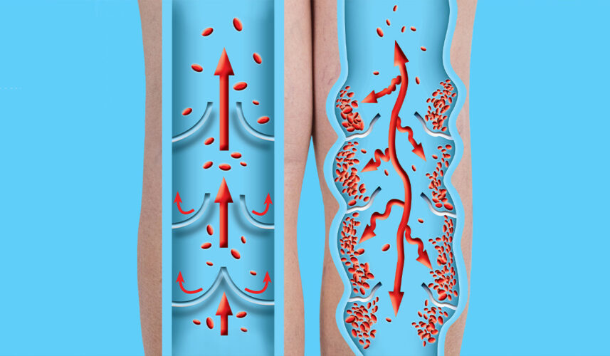
It is worth thinking about your heart before symptoms of heart failure appear. As mentioned, the most significant factor leading to coronary artery narrowing is atherosclerosis. Cholesterol deposits accumulate in the form of so-called atherosclerotic plaques, first causing narrowing and ultimately closing the lumen of the artery. High cholesterol levels are primarily caused by an incorrect diet, which is rich in animal fats and low in vitamins and antioxidants.
Natural treatment of angina involves eliminating the factors contributing to the disease, such as:
The fact that younger and younger people complain about heart problems is also related to the high demands placed on people by the modern world. Trying to cope with all our responsibilities, we eat poorly due to lack of time, do not get enough sleep, smoke cigarettes, abuse alcohol, and take more and more medications. With such a lifestyle, the protective effect of estrogens may be insufficient.

Cardiovascular diseases develop secretly over the years. You can prevent them![]() and track them down with tests – blood pressure, cholesterol and triglyceride levels, glucose. To avoid them, take care of your heart from a young age.
and track them down with tests – blood pressure, cholesterol and triglyceride levels, glucose. To avoid them, take care of your heart from a young age.
The best option is long, medium-intensity exercise, performed systematically – every day for 15 minutes or 3 or 4 times a week for at least 30 minutes. It's worth changing your habits: taking the stairs more often, walking or cycling part of the way to work.
There is no place for fatty animal products high in cholesterol. Eat like people living in the Mediterranean – they have the lowest incidence of cardiovascular diseases in Europe. Eat seafood and olive oil (they contain omega-3 fatty acids that are valuable for the heart), fresh vegetables and fruits, cereal products, groats, and legumes (they are rich in fiber, which lowers cholesterol levels).
This menu helps you lose weight. It is important because each additional kilogram forces the heart to work harder, and abdominal obesity is a health hazard.
Stress leads to excessive production of catecholamines (organic chemicals that include, for example, adrenaline), which have a negative effect on the heart. They lead to an acceleration of the heart rate, an increase in blood pressure, a slight dilation of the coronary vessels, and consequently overload the heart muscle. It is worth limiting exposure to stress factors and learning to fight stress. Known methods include Schultz's autogenic training, Jacobson's training, visualization, yoga, and meditation.
Sleep allows the body to regenerate after a day's effort, but is also a factor in recovery. Sleep deprivation contributes to endothelial dysfunction, increased activity of pro-inflammatory factors, increased oxidative stress, and many other pathological conditions that lead to changes in the vessels, heart, and kidneys. Their standard and dominant feature is hypertension. Poorer sleep quality may, therefore, increase the risk of cardiovascular diseases and promote the progression of chronic kidney disease. Sleep needs in humans depend on age. Adults should sleep 7-9 hours a day.