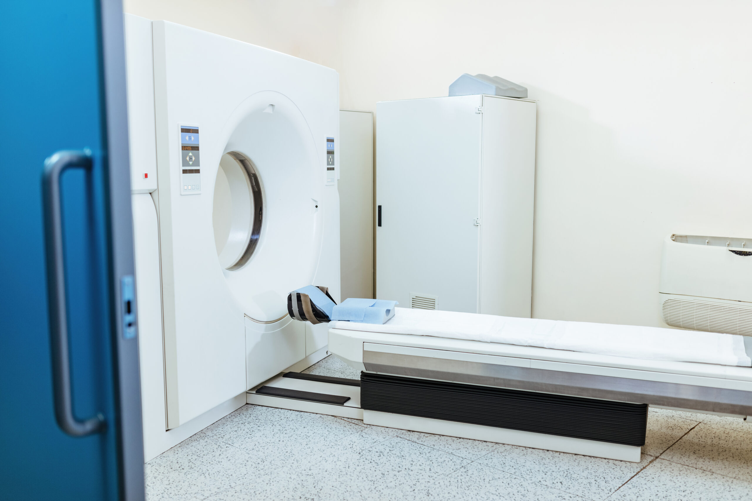
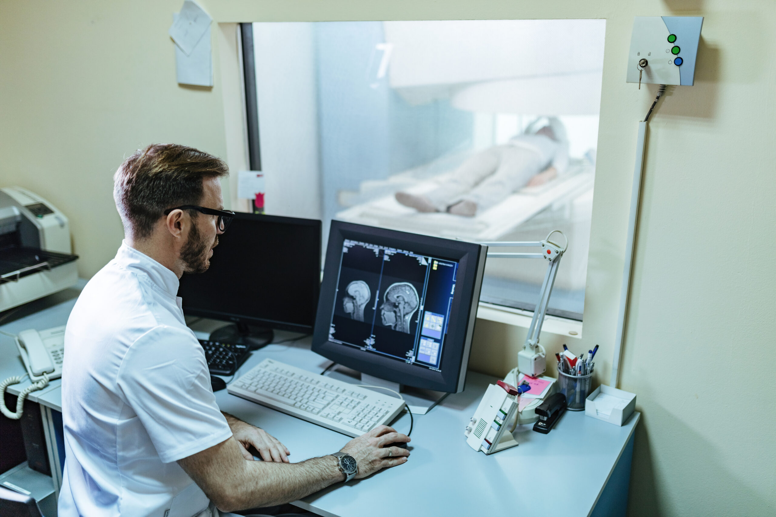
Magnetic resonance imaging (MRI) is a non-invasive diagnostic tool that helps identify various pathological changes within the body. This imaging procedure delivers cross-sectional pictures of internal organs, allowing them to be displayed in any desired plane. An MRI can often be beneficial if a computed tomography (CT) scan does not yield the expected results.

Magnetic resonance imaging![]() can be used to scan the entire body. Tissues covered by bone structure are particularly recommended for imaging with this method. These include the brain with the spinal cord (central nervous system), the heart, muscles, subcutaneous tissue of the limbs, and joints. The following are often assessed: the abdominal cavity, pelvis, bile ducts, and urinary system.
can be used to scan the entire body. Tissues covered by bone structure are particularly recommended for imaging with this method. These include the brain with the spinal cord (central nervous system), the heart, muscles, subcutaneous tissue of the limbs, and joints. The following are often assessed: the abdominal cavity, pelvis, bile ducts, and urinary system.
Magnetic resonance does not cause complications after the test compared to other radiological tests. It is safe enough to be used in pregnant women, but this condition should be reported before the test.
On the same day as your magnetic resonance imaging scan, you can usually eat, drink, and take any medicine as normal unless you're instructed otherwise.
When you arrive at the hospital, you must usually fill out a questionnaire about your health and medical history. This information helps the medical staff ensure you have a safe scan.
As the MRI scanner creates powerful magnetic fields, removing any metal things![]() from your organism is necessary. These may be:
from your organism is necessary. These may be:
Avoid bringing valuable items to the scan. Instead, store them in a secure locker.
Depending on the body part being scanned, you may need to wear a hospital gown. If that is not the case, the individual should wear clothes without metal zips, buttons, underwire, belts, or buckles.
Depending on the kind of test and the area being scanned, the examination![]() can be completed without preparation or requires a 2-hour fast. This time may be longer, especially if the test concerns the digestive tract or bile ducts.
can be completed without preparation or requires a 2-hour fast. This time may be longer, especially if the test concerns the digestive tract or bile ducts.
If general anesthesia is used, you must fast for 6 hours before the test. The urinary bladder should be moderately full if the test involves the pelvis. On the day of the test, take your medicines with a small amount of water as you normally do.
You should have a breast test between the 6th and 13th day of your cycle, even if you are using contraception. If you are taking hormone replacement therapy, the specialists recommend to complete the examination four weeks after discontinuing it.
You should also bring documentation from previous imaging studies (e.g., ultrasound, X-ray, CT scan) to the examination. Comparing previous studies often allows for a more accurate diagnosis.
The area being scanned should be clean and free of cosmetics, the composition of which can also generate distortions of the magnetic field. This applies particularly to make-up and hairspray while having a head examination.
Arrive for your MRI at least 30 minutes early. This gives you time to ask questions about the scan and to complete a questionnaire about your general health, past surgeries, and any metal objects in your body. Any metal objects, magnetic cards, and jewelry should be left in the changing room, even those in a part of the body far from the examination area. In contrast, those undergoing outpatient scans may have to wait a few days for their results.
The examination is lengthy. Depending on which body part is being imaged, the patient may stay in the tube for about 20 to 60 minutes.
Sometimes, it is necessary to perform more than one examination (for example, to diagnose all sections of the spine – cervical, thoracic, and lumbar).

The duration of the MRI scan is longer for many patients because they are completely immobilized and cut off from external stimuli. Some people, especially those suffering from claustrophobia, do not feel comfortable in such a tight space.
Especially since it is warm in the tube, and the sounds coming from the device are not the most pleasant. They are mechanical, loud, and sometimes unexpected – so they can be scary. Nevertheless, after prior mental preparation, the entire procedure can be completed without significant emotions.
MRI is a highly specialized examination, which is why there are many indications for magnetic resonance imaging. As mentioned above, magnetic resonance imaging can be used to examine any part of the body. Magnetic resonance imaging is indicated in the diagnostics of almost all systems. You can perform it in the case of:
Although MRI is a very accurate test for detailed diagnostics, not everyone can use it. There are numerous contraindications![]() to resonance imaging. These include:
to resonance imaging. These include:
In the above situations, most clinics should refuse to perform the test. In addition, a broad range of circumstances constitute relative contraindications to performing magnetic resonance imaging, which means that the doctor decides on the possibility of placing the patient in a tube each time. The obstacles that may stand in your way include:
Patients who have undergone heart surgery should not undergo MRI for some time – the waiting period is usually a dozen or so weeks.
Individual laboratories approach whether pregnancy is a contraindication to MRI differently. Some of them examine prior qualifications; for others, they do not. This is a problem that could exclude the patient from the examination.
If contrast is to be used, the criterion determining admission to diagnostics is the creatinine level. A separate issue is the aforementioned claustrophobia and other more or less serious anxiety disorders.
They are not a contraindication to the examination but may imply the need for a special procedure.
Magnetic resonance imaging (MRI) is generally regarded as a safe procedure![]() . The technique employs a magnetic field ranging from 0.5 to 3.0 Tesla. Studies have failed to show any harmful effects of MRI. However, it’s advisable to postpone MRI during the first trimester of the pregnancy
. The technique employs a magnetic field ranging from 0.5 to 3.0 Tesla. Studies have failed to show any harmful effects of MRI. However, it’s advisable to postpone MRI during the first trimester of the pregnancy![]() .
.
However, patient safety can be compromised if metal objects are inside the body, such as metallic foreign bodies, vascular clamps from previous surgeries, or various stimulators. This is especially true if these devices are powered on, as the MRI may disrupt their function. Thus, the existence of these metal elements is a contraindication for the process.
The powerful magnetic field made during the scan can move the sensitive to its effects objects that are even profoundly and firmly in the body.
Most implants in the body do not cause problems, but if they are close to the scanned area, they can distort the magnetic field and lower the quality of the images.
The exam typically takes 20 to 60 minutes, and the patient lies in a narrow chamber where crackling sounds can be heard. Because of this, people with claustrophobia![]() or those who cannot stay still may be unable to complete the exam, as their movements can create artifacts in the images.
or those who cannot stay still may be unable to complete the exam, as their movements can create artifacts in the images.
In many cases, a contrast agent is administered intravenously, so individuals with hypersensitivity to gadolinium contrast agents are advised against this examination. It's worth noting that allergic reactions to MRI contrast agents are generally milder and less frequent than those related to iodine contrast agents used in computed tomography (CT) scans.

A contrast agent![]() is a substance given through an injection or orally. It helps improve the quality of images taken during scans. For example, using a contrast agent during an MRI makes specific tissues show up better.
is a substance given through an injection or orally. It helps improve the quality of images taken during scans. For example, using a contrast agent during an MRI makes specific tissues show up better.
We often use gadolinium agents in MRIs. They are generally safer than iodine agents used in CT scans, as they are less likely to cause severe allergic reactions. In some cases, doctors may even give them to pregnant women if necessary.
The doctor or radiologist makes the choice to use a contrast agent. In some conditions, like certain rheumatological diseases or suspected strokes, using a contrast agent can greatly help with diagnosis. However, the scan results can be good enough in other situations without it.
Contrast agents can cause acute side effects![]() (within 1 hour of administration), those include:
(within 1 hour of administration), those include:
These are usually responses resulting from hypersensitivity – allergy-like. Patients who have previously experienced a similar reaction or are burdened with asthma and atopy are most at risk of side effects. Most often, the side effects are mild, short-term, and transient.
Late side effects after the administration of gadolinium-based contrast agents include nephrogenic systemic fibrosis. Symptoms may occur up to several months after contrast administration. They usually concern the lower limbs and include:
As a result, thickening and fibrosis of the skin, sometimes contractures of the limbs, and even fibrosis of the internal organs may occur (patients with impaired kidney function and undergoing dialysis are at risk of this side effect).
It should be noted that this severe side effect is rare and mainly affects people suffering from severe kidney disease. To minimize the chance of side effects, the lowest possible dose of contrast is used in all individuals. Patients also have their blood creatinine levels measured before the test to assess kidney function.
After the contrast test, the patient is left under observation for about 30 minutes due to the possibility of an acute side effect. Then, they go home with instructions to drink plenty of fluids to help the contrast agent be excreted in the urine.
An MRI scan is usually an outpatient procedure, so the patient doesn't need to stay overnight in the hospital.
After the examination, the individual can resume normal activities immediately. However, if a specialist uses a sedative, a friend or relative should take the patient home and remain with them for the first 24 hours. After receiving a sedative, the patient should not drive, operate heavy machinery, or drink alcohol for 24 hours.
A radiologist will review the MRI scan and may discuss it with other specialists. Therefore, it's unlikely that results will be available immediately.
If there is pain or any unusual symptom following the exam, the patient should contact the physician.