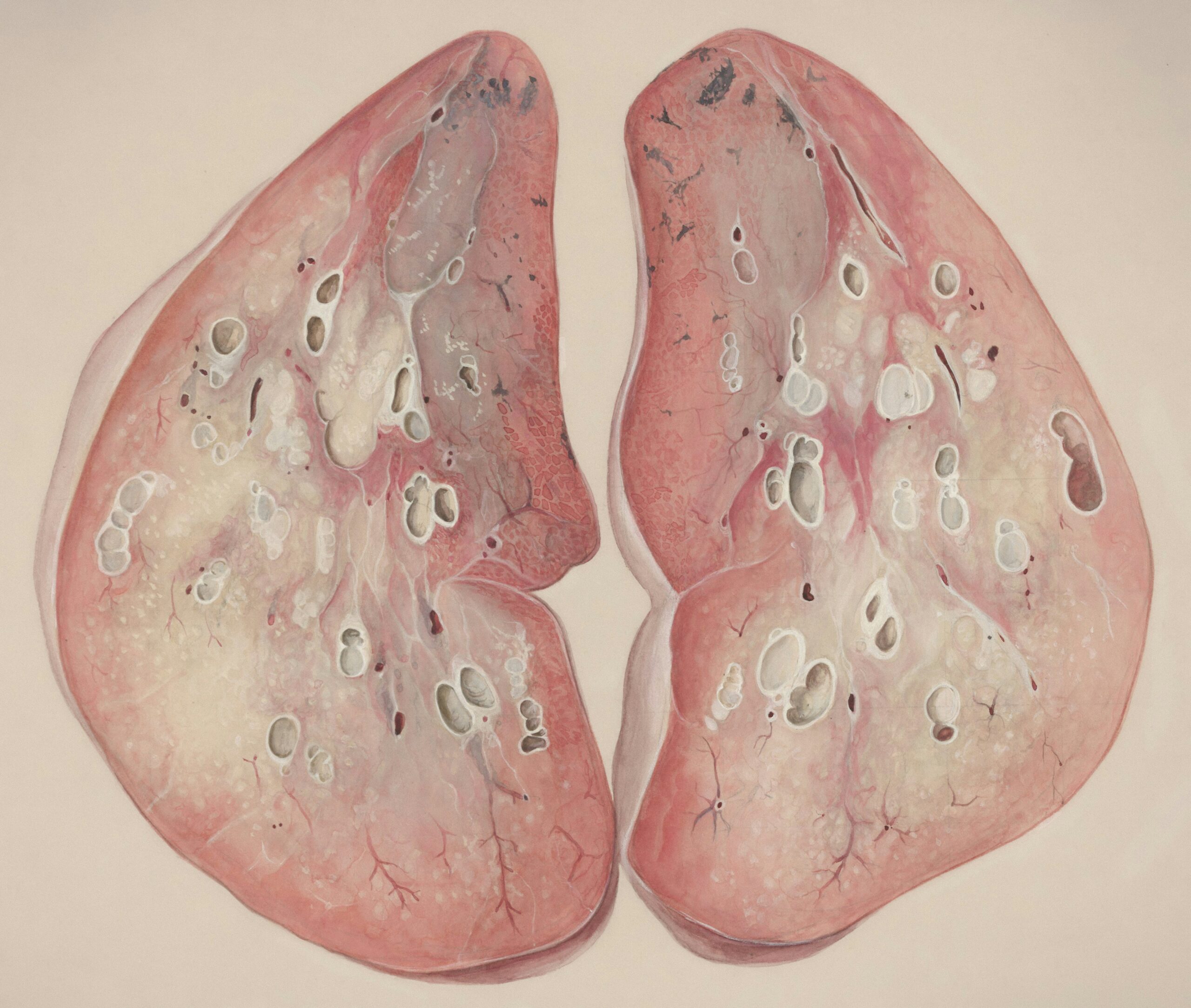
This condition is called pleural effusion, and it is explained as when too much fluid collects in the small space between the rib cage and lungs. In most circumstances, the pleural space is filled with a bit of fluid that gives the lungs the freedom of perfect movement in the entire process of respiration.
The other end of this (excessive fluid intake) can become a zone of conflict. That is nothing but an expression of the fact that the body is undergoing a struggle with some other problem. As mentioned in the statement, it can also be caused by pathogens, lung and heart diseases, or cancer.
Pleural effusion is the root cause of the problem, which in turn impairs the patient's lungs and makes breathing easier and more comfortable. The primary objectives of treating pleural effusion include identifying the causes of pleural effusion and experimenting with effective methods for fluid drainage.

How significant is the percentage of patients who usually get sick with pleural effusion? But, you should recognize that pleural effusion isn't just the least common of all, even though it's common among hardy old adults.
The data provided by the National Heart, Lung, and Blood Institute (NHLBI) shows a strong positive correlation between smoking and new cases of pleural effusion (only in the US), which accounts for more than a million new cases every year. Pleural effusion is a common illness that has been the cause of distress in the lives of people across the globe from time immemorial.
Some of the affected people will be those who have undergone surgeries as well, those who are suffering from heart diseases and pneumonia, and some cancer patients will be the people who will face these after the operation. Even though it exists in all age groups, habitually feeble elderly ones, along with individuals suffering from diseases such as heart, lung, and kidney problems, are more likely to develop it. The major causes of pleural effusion include the patient's advanced age and general health.
The causes of pleural effusion and the people affected by it vary. The most likely scenario is when it appears as an inability to control a fluid buildup that has accumulated over time; thus, heart failure is the key factor in this situation. Because of the au's near impossibility to pump blood from the lungs at a low rate, the fluid gets stuck in the pleural space. Thus, the pleural space becomes overgrown and consequently remains distended.
For example, pleural effusion can be caused by diseases, such as pneumonia, that trigger the immune system to manufacture an oversized quantity of fluid and inflammation that wick into the pleural membrane.
This is another cause of pleural effusion. There is also regrettable involvement of cancer, which might be, for instance, lymphoma, breast cancer, and lung cancer. When these cancers are localized in the pleural space or depleted the lymphatic system, the fluid is generated.
On the contrary, problems such as renal failure and cirrhosis might indirectly lead to pleural effusion through the disturbance of fluid balance. Autoimmune conditions, such as multiple sclerosis, rheumatoid arthritis, and lupus, among others, are associated with the activation of unwanted inflammation, hence the causative of pleural effusion. Furthermore, the contributory, albeit rare, causes are operation problems, thorax damage, and treatment options, like the use of certain medications that cause inflammation in the pleural space.
Symptoms of pleural effusion may appear gradually or suddenly depending on whether the fluid accumulation is fast and the underlying problem is present.
The most likely symptom of excessive airway content is the feeling of a short breath, which can initially be moderate but can be life-threatening when there is a great amount of oxygen in the air. Nearly half of all patients describe a tight chest as the predominant symptom of not breathing correctly.
Moreover, pleural effusion can be the source of various chest symptoms, which are usually recognizable if the patient is breathing in or coughing a lot and may be regularly felt.
On the other hand, fever and shivering are the two most approved symptoms that may accompany the infection if it is the required cause. The most prevalent symptom of it is joined by a lack of appetite and the loss of body weight, especially when the condition is cancer or other chronic diseases.
Fatigue is also a component of the disorder. Some people may suffer the discomfort of a “dry” cough![]() or not notice any additional symptoms at all. Nevertheless, there are people whose symptoms have become so serious that they must visit a medical doctor.
or not notice any additional symptoms at all. Nevertheless, there are people whose symptoms have become so serious that they must visit a medical doctor.

If the pleural effusion is not treated, the problems may worsen. One of the earliest manifestations of the disease is the constant buildup of fluid in the body, which causes the body to become compressed and makes breathing more difficult.
The tension of this situation is that the human respiratory system cannot get the necessary oxygen due to the restrictive lungs, which consequently leads to the discomfort felt by the body. Infection with pus in the pleural fluid is another important issue; the name of this condition is empyema, and it causes the fluid to be invaded by pus.
Empyema![]() is not just an irritation; although it is a risk, it can evolve into an infection that results in blood poisoning. Inflammation, thickening of the pleural membrane, and pleural effusion are the three pleural issues that occur the most frequently. The thickening of the pleura usually comes from long-lasting inflammation even though it can sometimes happen due to fluid evacuation.
is not just an irritation; although it is a risk, it can evolve into an infection that results in blood poisoning. Inflammation, thickening of the pleural membrane, and pleural effusion are the three pleural issues that occur the most frequently. The thickening of the pleura usually comes from long-lasting inflammation even though it can sometimes happen due to fluid evacuation.
Pleural effusion, which is a recurring phenomenon, could be a peculiar problem, especially when the condition is persistently caused by the underlying disease. Nevertheless, this may lead to countless fluid removal procedures as well as other treatments in cases where the region becomes particularly invaginated due to the fluid.
It is a physical examination that reveals the presence or absence of pleural fluid. In such instances, physicians may place their ears on the chest to listen to the sounds of breath or dull noises. At first, the swelling shows that you are keeping water in your body. Nevertheless, they need to be more competent in describing the type of water being preserved, that is, corporeal or renal, or specifying the causes of water retention.
The chest X-ray![]() is the most primary diagnostic test of the human body and was the first test to be introduced. It is helpful in positioning and quantifying the fluid in the chest area. It is true that the liquid is moving from one side to the other while the person is lying down; thus, the pleural space does not consist of only solid tissue. Still, X-ray data only supplies some of the information you need.
is the most primary diagnostic test of the human body and was the first test to be introduced. It is helpful in positioning and quantifying the fluid in the chest area. It is true that the liquid is moving from one side to the other while the person is lying down; thus, the pleural space does not consist of only solid tissue. Still, X-ray data only supplies some of the information you need.
An ultrasound is quite accurate as it definitively and unambiguously identifies the fluid location, which is crucial in a thoracentesis process to drain the fluid. Ultrasonography is also an efficient way to find small amounts of fluid trapped in specific locations. It can also be applied to aspirate this fluid for diagnostics.
A CT scan is imaging you can perform to get more information about a patient. Suppose the effusion is used in a neoplasm or a critical disease. In that case, a CT scan can help medical personnel obtain additional information, namely, pleural thickening, suspicious growths, or even signs of infection. This type of testing has been proposed to be applied in cancer diagnostic processes since this analysis not only shows how far the cancer has spread but also allows physicians to choose the most effective treatment among available choices.
A thoracentesis![]() is the process of retrieving the required fluid by sticking the needle through the chest wall and into the pleural cavity. Apart from providing symptomatic relief through fluid removal, it also allows further research that presents information such as protein level, glucose, and cell count in the body. One of those tests that reveal your type of leak is an important step in the problem you face.
is the process of retrieving the required fluid by sticking the needle through the chest wall and into the pleural cavity. Apart from providing symptomatic relief through fluid removal, it also allows further research that presents information such as protein level, glucose, and cell count in the body. One of those tests that reveal your type of leak is an important step in the problem you face.
This analysis involves acquiring information via laboratory testing. By interpreting the concentrations in substances like pH, glucose, and protein, it can be determined that the fluid is definitely inflammatory, infectious, or malignant. A few tests, such as cytology, cultures, and chemical composition assays, are involved in finding the causes, which, in return, helps in aiming at the treatment formulation.
In some occasions, the biopsy is the last test that will be needed to verify a cancer diagnosis that is indicated but not confirmed in the fluid analysis. That is one of the situations that may occur. In some cases, it is possible to obtain tiny bits of tissue by the use of a needle or a thoracoscopy procedure. This enables histological sections to be taken from pleural tissue and the test to be done for malignancy.

The main treatment method for pleural effusion consists of relieving the symptoms, collecting the fluid, and finding and treating the underlying cause so that it never recurs. Different treatment options can be available depending on the extent of the problem.
Other than being used to diagnose diseases, thoracentesis is also one of the most effective methods of swiftly providing symptom care. After the needle removes the fluid from the body, the patient can sometimes detect a sudden relief of pressure and a decrease in shortness of breath. An ultrasound is usually used to ensure that the correct spot is located to insert the needle.
Chest tube insertion is a regular treatment used often when there are large or occurring effusions. It is also largely used to contain dirty fluid or fluid trapped in pockets. The lungs are allowed to expand when a tube is placed; it is then instructed to drain continuously. However, this therapy is a great way to prevent the fluid from going back too soon, and it is very useful when there is an infection or a tumor involved.
One of the most common remedies for patients suffering from this lung condition is called pleurodesis. This procedure aims to cause the lung linings to stick together by injecting a chemical irritant, which is talc in most cases, into the pleural space. On the one side, pleurodesis is the most evident upside to being at the risk of a patient's suffering who released from conformity to frequent lectures is, (a) it performs, besides facilitating a change in the way that fluids may accumulate are removed, also the function of the main of the treatment solution.
In general, medications are used as an auxiliary therapy and especially in case of infectious and inflammatory conditions. If there is inflammation, antibiotics are administered, and diuretics will be helpful for patients who have heart failure or kidney issues, in addition to it being a prescription for the management of fluid retention.
The most severe scenarios and persistent effusions that do not respond to standard medications are the only indications of surgery. If the pleural layer is very thick and the lungs cannot expand properly, medical treatment of a pleurectomy (the excision of part of the pleura) or decortication (removal of fibrous tissue) might be considered.
The debut of sickness-related symptoms and the need for fluid management in patients whose diseases are protein-effective are the reasons you should allow them to decide between the dependency on treatment or the dependency on chronic effusions, and they should also be directed to you to explain their needs to you. Patients can do their daily initial draining efficiently in the house using these catheters, sometimes skipping many journeys to the hospital.