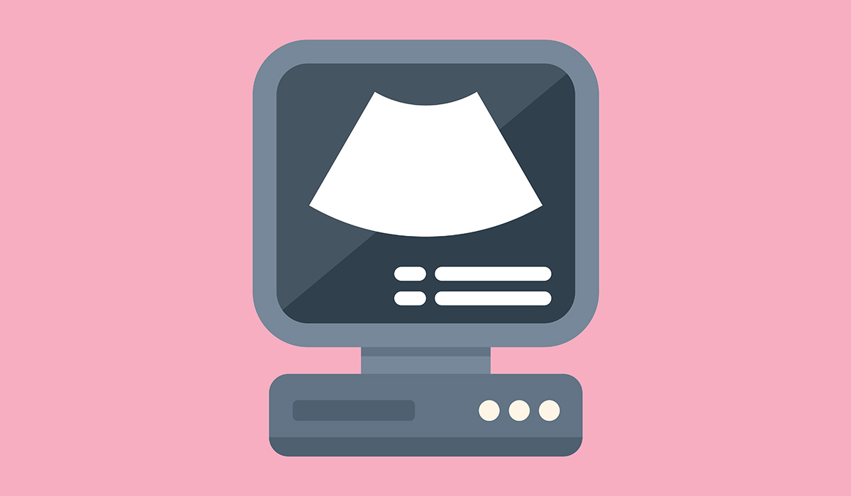
An echocardiogram is an ultrasound imaging examination of the heart. It is based on the emission and recording of ultrasonic waves. The acoustic wave generated by the device reaches deep into the tissues and, depending on the tissue bounces off them. The ultrasonic signal is converted into electric energy transmitted to the ultrasound computer, which shapes the appropriate image.
An echocardiogram allows you to determine the heart structure, size, and functioning of the heart valves and to measure the work of the organ.
Echocardiography![]() is a non-invasive examination. The specialist can perform transesophageal or transthoracic echocardiogram, but there are also other types (which we will discuss later).
is a non-invasive examination. The specialist can perform transesophageal or transthoracic echocardiogram, but there are also other types (which we will discuss later).
For a transthoracic examination, the patient removes their clothes from the waist. They lie on a couch on their back or left side. The specialist spreads a transducer lubricated with a certain gel to the chest. The transducer is moved to the appropriate places to obtain the right image. The examination lasts about 15-30 minutes depending on the detected abnormalities and the patient's general condition.
Transesophageal echocardiography is performed with a thin ultrasound probe inserted into the esophagus. It is directly adjacent to part of the heart, so reducing the distance allows for a more accurate image and data collection. Before the probe is inserted, the patient receives local anesthesia and, if necessary, a sedative.
Applications of echocardiography![]() include:
include:
Modern echocardiographic technology allows for non-invasive, safe, and accessible diagnostics. Recent developments have made it possible to conduct heart ultrasounds at the patient’s bedside and home with pocket devices. However, these compact devices remain less popular due to their high cost.
Echocardiography utilizes three imaging modes: one-dimensional, two-dimensional, and three-dimensional. It can be performed using transthoracic, transesophageal, and intraoperative techniques.
The most basic mode is one-dimensional, which allows healthcare providers to observe the movement of selected structures along a single line. This mode is particularly beneficial in cardiology for evaluating heart valves and vessel elasticity.
The two-dimensional mode, the most widely used in medicine, provides a broader view of surfaces and enables numerous calculations crucial for diagnosis.
Three-dimensional imaging, or 3D, is a more recent advancement that offers depth perception, proving especially advantageous in preparations for cardiac surgery.
Transthoracic echocardiography![]() is the most fundamental and widely utilized imaging test in contemporary cardiology. This technique allows for the heart and vessels visualization from various angles, including parasternal, apical, suprasternal, and substernal views. The specific projection selected depends on the structures the examining physician aims to assess.
is the most fundamental and widely utilized imaging test in contemporary cardiology. This technique allows for the heart and vessels visualization from various angles, including parasternal, apical, suprasternal, and substernal views. The specific projection selected depends on the structures the examining physician aims to assess.
In conjunction with the echocardiogram, Doppler imaging is routinely employed during the procedure. The techniques allow you to obtain image depth, which is especially useful before cardiac surgery.
Modern echocardiographic devices enable non-invasive, safe, and accessible diagnostics. Technological development has made it possible to perform heart ultrasound at the bedside and at home using so-called pocket devices, which are not very popular due to their high price.
The specialists perform transesophageal echocardiography![]() by inserting a very thin ultrasound probe into the esophagus directly adjacent to the left side of the heart. By decreasing the space between the probe and the observed objects, it is possible to obtain an image with a much higher resolution, i.e., accuracy. The projections used in transesophageal echocardiography are the low, middle, and high esophageal projections and the aortic and transgastric projections.
by inserting a very thin ultrasound probe into the esophagus directly adjacent to the left side of the heart. By decreasing the space between the probe and the observed objects, it is possible to obtain an image with a much higher resolution, i.e., accuracy. The projections used in transesophageal echocardiography are the low, middle, and high esophageal projections and the aortic and transgastric projections.
Thanks to the miniaturization of diagnostic equipment, it is possible to use the benefits of ultrasound also during the surgical procedure. In heart diseases, intraoperative echocardiography![]() is limited to cardiac surgery. This examination is used only for specific indications.
is limited to cardiac surgery. This examination is used only for specific indications.
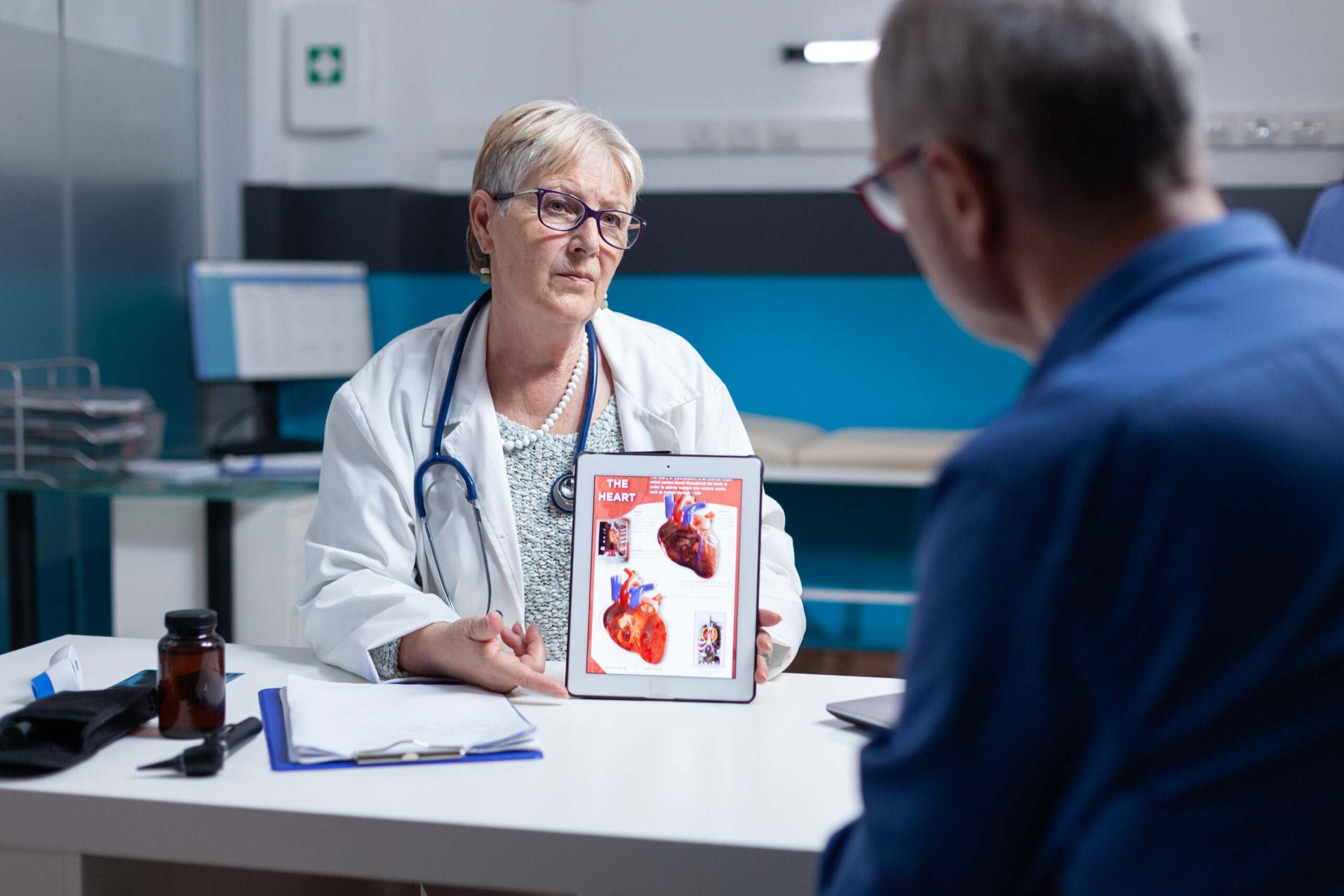
Before transesophageal echocardiography, you need to prepare![]() , the doctor thoroughly explains to the patient what the examination involves so that the patient can give informed consent to undergo it. The specialist will also question the patient about their health situation and whether they have any of the following:
, the doctor thoroughly explains to the patient what the examination involves so that the patient can give informed consent to undergo it. The specialist will also question the patient about their health situation and whether they have any of the following:
For the examination, it is necessary to remove removable dentures and insert a cannula into a vein to administer drugs. During the examination, a pulse oximeter is placed on the patient's finger to monitor blood oxygenation and a monitored ECG from one lead. Occasionally it is essential to administer oxygen intranasally.
The patient should not eat or drink for at least 4 hours before the examination and up to 2 hours after the examination.
An echocardiogram can be performed![]() in a hospital, clinic, or specialist's office.
in a hospital, clinic, or specialist's office.
It can also be performed in a hospital room at the patient's bedside. The patient should remove clothing and jewelry from the waist up. Transthoracic echocardiography, Doppler echocardiography, and stress echocardiography are performed by specialized sonographers. Transesophageal echocardiography is performed by a cardiologist.
The patient may have an intravenous line to administer medication during the test. A contrast agent may also be given intravenously to enhance the clarity of heart images, especially useful when it's challenging to get clear visuals – such as in cases of obesity or chronic lung disease.
During transthoracic echocardiography with Doppler![]() , the patient is positioned on their left side, with electrodes attached to their arms and legs to monitor the heart rhythm throughout the test. The doctor applies a small amount of a special gel to the patient's chest, enhancing the transmission of ultrasound waves. The ultrasound head is placed on the patient's body and the doctor slowly moves it along the chest wall. The device sends ultrasound waves and then receives echoes as they bounce off various internal organs.
, the patient is positioned on their left side, with electrodes attached to their arms and legs to monitor the heart rhythm throughout the test. The doctor applies a small amount of a special gel to the patient's chest, enhancing the transmission of ultrasound waves. The ultrasound head is placed on the patient's body and the doctor slowly moves it along the chest wall. The device sends ultrasound waves and then receives echoes as they bounce off various internal organs.
Then the received signals are sent to a monitor, which processes them into images of the heart's components. The room is usually darkened so that the pictures on the monitor are easier to see. The specialist may ask the patient to lie still, slowly inhale and exhale, and hold their breath. The head is moved in different directions to obtain different cross-sections of the heart. The examination usually lasts from 30 to 60 minutes. After the examination, the gel is wiped off and the electrodes are peeled off.
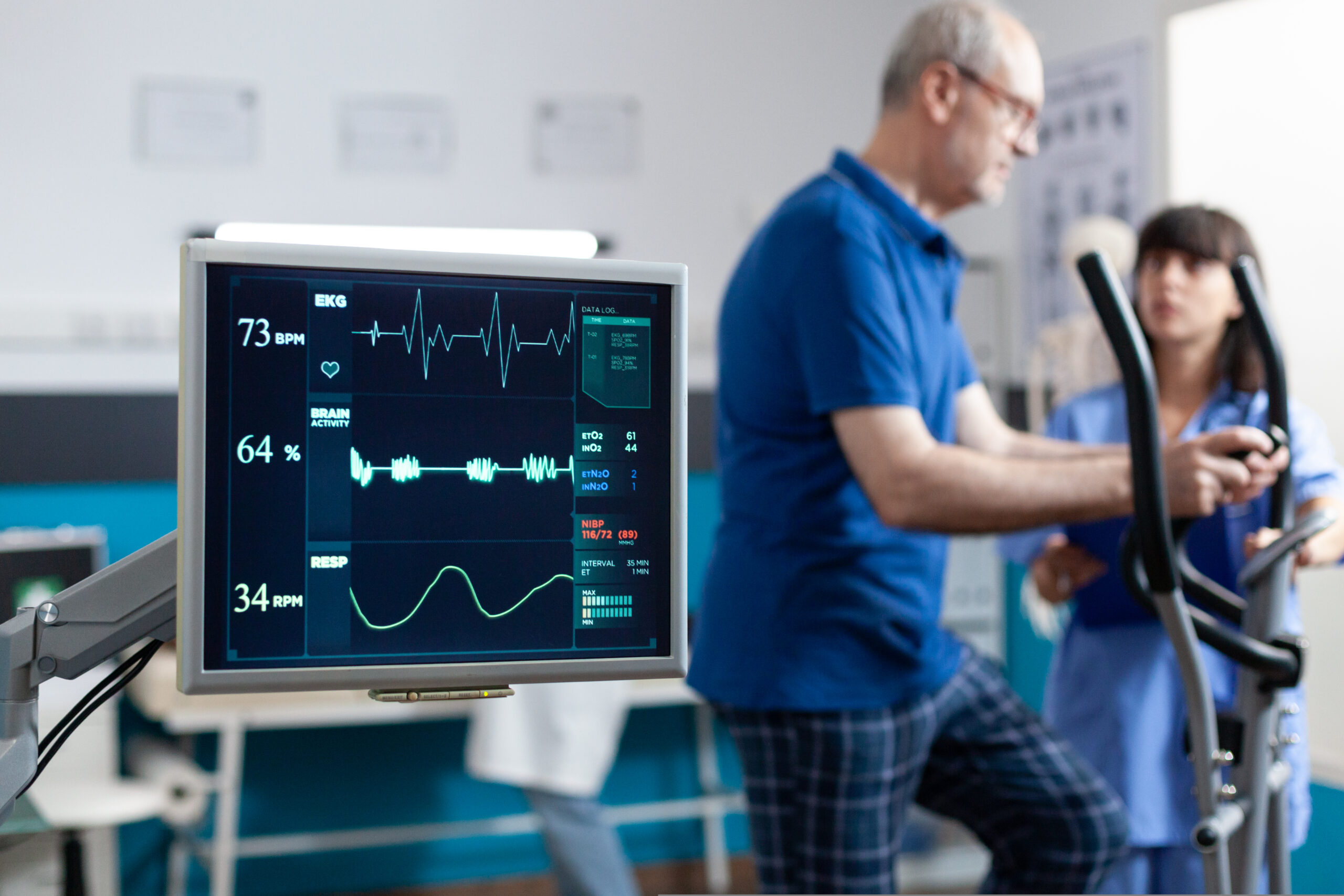
An exercise stress echo![]() evaluates the heart’s function when it’s beating quickly. The patient creates this “stress” by exercising on a treadmill or bicycle.
evaluates the heart’s function when it’s beating quickly. The patient creates this “stress” by exercising on a treadmill or bicycle.
In some situations, rather than relying on physical exercise, a medication![]() is used to place stress on the heart. During this examination, the patient lies on their back or side while the initial echocardiogram is conducted.
is used to place stress on the heart. During this examination, the patient lies on their back or side while the initial echocardiogram is conducted.
Next, an intravenous line is set up, and medicine is introduced. This medication boosts both the frequency and strength of the heartbeat. Typically, the peak heart rate is achieved about 15 minutes after medication is administered. While this occurs, another echocardiogram is performed. The doctor might ask the patient to remain still, breathe in and out slowly, and then hold their breath.
A doctor provides the medicine into the IV and the technician resumes to move the transducer to obtain echo images. The medicine forces the heart to respond as if you were training. After the test, the healthcare provider pulls out the IV from the patient's arm. The heart rate will return to standard within five to 10 minutes.
A transesophageal echocardiogram (TEE) uses sound waves to make images of the heart. It differs from other echocardiogram forms or ultrasounds because it takes photos from within the organism, rather than outside.
The specialist uses a long, thin tube called an endoscope to carefully navigate a small transducer down the throat and esophagus. The transducer is an instrument that produces sound waves – they bounce off the various heart areas, making echoes. The transducer then transmits these echoes to a computer that creates images. These photos demonstrate the heart's structure and function in excellent detail.
The most important contraindication to transesophageal echocardiography is the patient's lack of consent to the test. TEE should not be performed in patients with esophageal diseases or bleeding diathesis. Patients who are HIV or hepatitis B carriers should report this fact to their doctor.
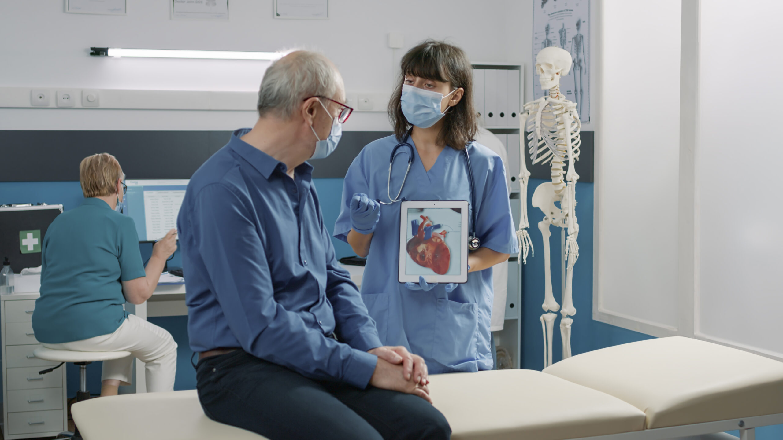
Echocardiography is used in the prevention and diagnosis of many diseases![]() . It allows for the detection of anatomical defects in the structure of the heart, various irregularities in the functioning of the organ, and damage caused by a heart attack. Thanks to ultrasound, the doctor can assess the size of the heart muscle and the volume of the pericardial sac.
. It allows for the detection of anatomical defects in the structure of the heart, various irregularities in the functioning of the organ, and damage caused by a heart attack. Thanks to ultrasound, the doctor can assess the size of the heart muscle and the volume of the pericardial sac.
In people with diagnosed heart disease or who are at risk, it is significant to have regular echocardiography (at least once a year).